. 2024 Sep 12;15(1):7898.
doi: 10.1038/s41467-024-51745-4.

A single-domain antibody targeting factor XII inhibits both thrombosis and inflammation

Affiliations

A single-domain antibody targeting factor XII inhibits both thrombosis and inflammation

Pengfei Xu et al. Nat Commun. .

Abstract

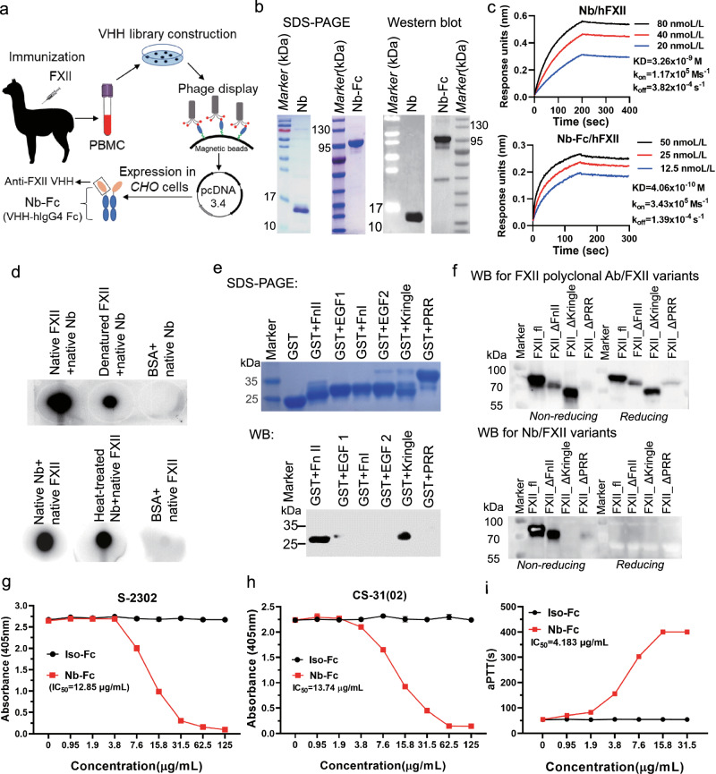
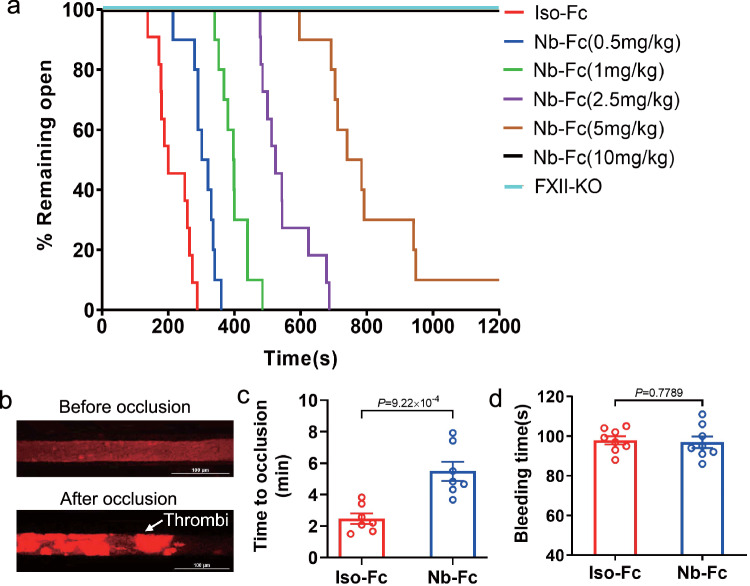
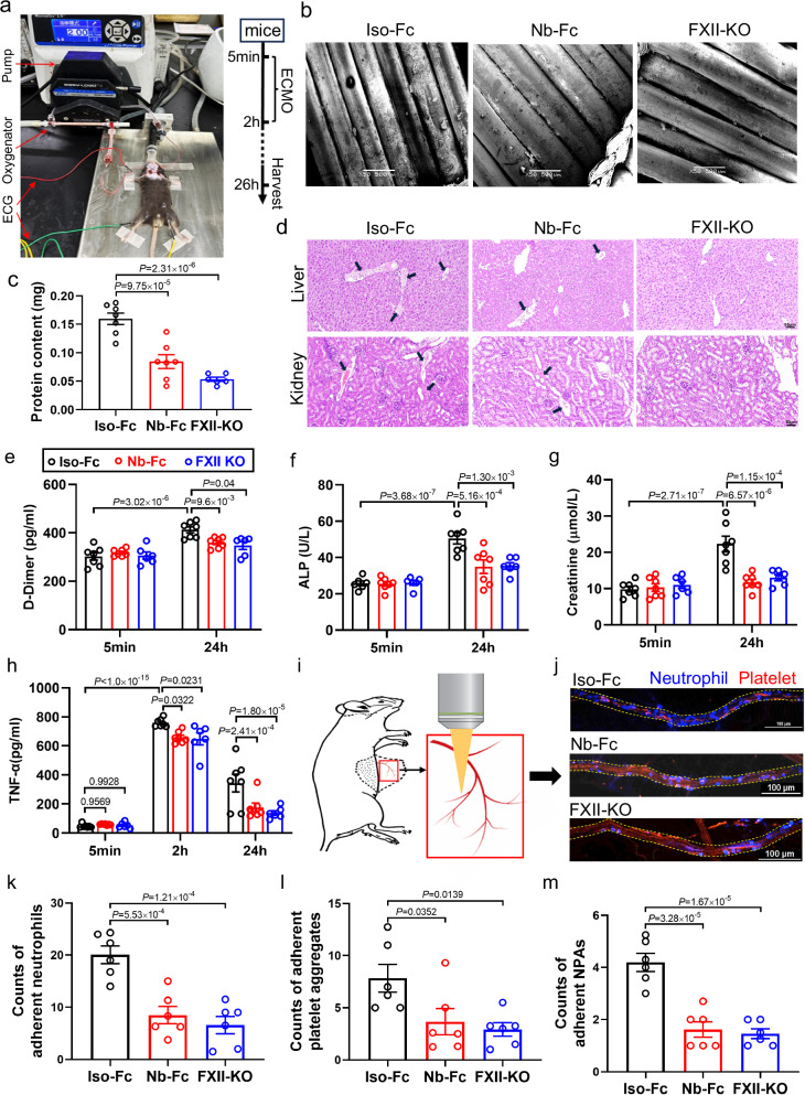
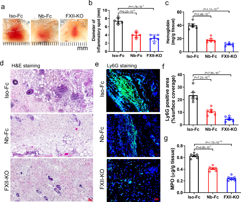
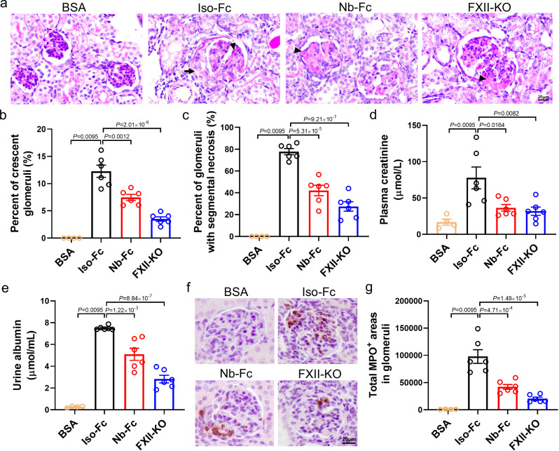
Factor XII (FXII) is the zymogen of the plasma protease FXIIa that activates the intrinsic coagulation pathway and the kallikrein kinin-system. The role of FXII in inflammation has been obscure. Here, we report a single-domain antibody (nanobody, Nb) fused to the Fc region of a human immunoglobulin (Nb-Fc) that recognizes FXII in a conformation-dependent manner and interferes with FXIIa formation. Nb-Fc treatment inhibited arterial thrombosis in male mice without affecting hemostasis. In a mouse model of extracorporeal membrane oxygenation (ECMO), FXII inhibition or knockout reduced thrombus deposition on oxygenator membranes and systemic microvascular thrombi. ECMO increased circulating levels of D-dimer, alkaline phosphatase, creatinine and TNF-α and triggered microvascular neutrophil adherence, platelet aggregation and their interaction, which were substantially attenuated by FXII blockade. Both Nb-Fc treatment and FXII knockout markedly ameliorated immune complex-induced local vasculitis and anti-neutrophil cytoplasmic antibody-induced systemic vasculitis, consistent with selectively suppressed neutrophil migration. In human blood microfluidic analysis, Nb-Fc treatment prevented collagen-induced fibrin deposition and neutrophil adhesion/activation. Thus, FXII is an important mediator of inflammatory responses in vasculitis and ECMO, and Nb-Fc provides a promising approach to alleviate thrombo-inflammatory disorders.


Conflict of interest statement

M.W. is a co-founder of NINGBO COMGEN BIOTECH CO., LTD. The remaining authors declare no competing interests.

Figures

Fig. 1
Fig. 1. Generation and characterization of FXII-specific nanobody (Nb) and its Fc construct variant fused to the Fc of human immunoglobulin G4 (Nb-Fc).
a Schematic representation of the strategy used to identify FXII-specific nanobodies and to design nanobody-Fc fusion after immunizing an alpaca with human FXII. b Characterization of E. coli BL21-expressed Nb and Chinese Hamster Ovary (CHO) cell-expressed Nb-Fc by SDS-PAGE (left panel) and Western blot (right panel). c Binding affinity of Nb or Nb-Fc to human FXII determined by kinetic assays using the Fortebio Octet System. d Dot blot assay to analyze the interaction of native or heat-treated FXII and Nb as indicated. BSA was used as control. e, f FXII epitope mapping. e FXII domains were individually expressed using E. coli BL21, separated by SDS-PAGE under reducing conditions and visualized by Brilliant Coomassie Blue (upper panel), and probed for Nb binding by Western blot (lower panel). f Full-length FXII (FXII_fl) and FXII mutants with indicated deleted domains (FXII_ΔFnII: deletion of FnII; FXII_ΔKringle: deletion of the Kringle domain; FXII_ΔPRR: deletion of the C-terminal part of PRR) were expressed in HEK 293 cells. Binding capacity of a polyclonal anti-FXII antibody as control (upper panel) and Nb (lower panel) to the mutants was evaluated by Western blot under both reducing and non-reducing conditions. g Effects of increasing concentrations of Nb-Fc on ellagic acid (EA, 4 µg/ml)-induced FXII activation using the FXIIa-specific substrate S-2302 (final concentration: 0.8 mM). Isotype control antibody (Iso-Fc) was used as control. h Effects of Nb-Fc on plasma prekallikrein (PK) activation. FXII was pretreated with Nb-Fc, followed by the addition of EA (final concentration: 4 μg/mL) and PK (6.25 μg/mL), then the chromogenic substrate CS-31(02) was used to evaluate the enzymatic activity of activated PK. i Effects of Nb-Fc on activated partial thromboplastin time (aPTT). Citrated human plasma was incubated with increasing concentrations of Nb-Fc (0–31.5 µg/ml) for 5 min prior to aPTT measurements. Experiments in b, df were independently performed twice with consistent results. See the Methods for details. Source data are provided as a Source Data file.
Fig. 2
Fig. 2. Treatment with Nb-Fc inhibits thrombosis without affecting hemostasis in mice.
a Nb-Fc inhibited 7.5% FeCl3-induced carotid arterial thrombosis. C57BL/6J mice were intraperitoneally injected with Nb-Fc or isotype control, Iso-Fc (n = 7–11), and F12−/− mice were used as control (n = 7). Time to vascular complete occlusion was monitored. b, c Nb-Fc treatment inhibited cremaster arterial thrombosis. Mice were intraperitoneally injected with 1 mg/kg Nb-Fc. Subsequently, mice were injected intravenously with 2% rose bengal (dissolved in PBS, 50 mg/kg) and PE-conjuated anti-CD41 antibody (2 μg/mouse), followed by 565-nm laser illumination at the cremaster artery. Representative images of thrombus formation taken by intravital microscopy (b, scale bar: 100 μm). Time to complete occlusion in the presence of Nb-Fc or Iso-Fc (c, n = 7). d Nb-Fc treatment did not affect hemostasis in mice (n = 8). Tail bleeding time in mice treated with 10 mg/kg Nb-Fc or Iso-Fc. The mean ± SEM of all data points are shown (c, d). Unpaired two-sided Student’s t test was used. Source data are provided as a Source Data file.
Fig. 3
Fig. 3. FXII knockout or inhibition with Nb-Fc suppresses ECMO-related thrombosis and inflammation and ameliorates organ injury in mice.
a Photographic illustration of the mouse ECMO setup and the experimental procedure ECMO. The Iso-Fc group and the Nb-Fc group were both treated with heparin (400 U/kg). The Nb-Fc group also received coadministration of Nb-Fc (2 mg/kg), and FXII knockout mice were used as positive control, which were intravenously injected with heparin (400 U/kg) (n = 6, 7). b Oxygenator membranes of mouse ECMO were analyzed by scanning electron microscopy (SEM), scale bar: 500 μm. c Protein content on oxygenator membranes (n = 6, 7). d Representative pictures from H&E staining of liver or kidney. Arrows indicate areas with thrombi, scale bar: 50 μm. Plasma D-dimer (e), ALP (f), creatinine (g) of mice underwent 5-min ECMO (baseline) or mice after 24-h ECMO (n = 6, 7). h Plasma TNF-α of mice underwent 5-min or 2-h ECMO (baseline), and mice after 24-h ECMO (n = 6, 7). i Schematic of the mouse microcirculation detection by spinning disk confocal microscopy. After 24-h ECMO, the mice were anesthetized with pentobarbital sodium, BV421-anti-Ly6G antibody (2 μg/mouse) and PE-anti-CD41 antibody (2 μg/mouse) was injected to the mice intravenously. The abdominal skin was cut along the midline of the abdominal cavity. The abdominal skin (inside side up) was placed on the stage of the spinning disk confocal microscope. The adherent neutrophils (blue), platelet aggregates (red) or neutrophil-platelet aggregates (NPAs, blue & red) in the venules were detected by spinning disk confocal microscopy. j Representative pictures of adherent neutrophils, platelet aggregates and NPAs in venules. Scale bar: 100 μm. Counts of adherent neutrophils (k), platelet aggregates (l), NPAs (m) in venules (n = 6). Data are shown as mean ± SEM. One-way ANOVA with Dunnett’s multiple comparisons test (c, km). Two-way ANOVA with Sidak’s multiple comparisons (eh). Source data are provided as a Source Data file.
Fig. 4
Fig. 4. FXII knockout or inhibition with Nb-Fc ameliorates immune complex-mediated vasculitis in mice.
a Representative macroscopic findings of hemorrhage in ICV mice. Wildtype mice were intraperitoneally injected with Nb-Fc or Iso-Fc (2 mg/kg), and FXII knockout mice were used as positive control. After 30 min, the mice were subjected to BSA (75 µg/g) and anti-BSA antibody (60 µg/mouse). Skin tissues were harvested four hours post BSA and anti-BSA antibody treatment (n = 6). b Diameter of the skin hemorrhage spots (n = 6). c Quantification of tissue hemoglobin levels by ELISA (n = 6). d H&E staining and e immunofluorescence staining of the dermal hemorrhage spots using an antibody specific for Ly6G (marker for neutrophils). Scale bar: 50 μm. f Quantification of Ly6G positive (green) areas (n = 6). g The MPO contents of mice in each group (n = 6). Data are presented as mean ± SEM. One-way ANOVA Dunnett’s multiple comparisons test (b, c, f, g) was used. Source data are provided as a Source Data file.
Fig. 5
Fig. 5. FXII knockout or inhibition with Nb-Fc ameliorates ANCA associated systemic vasculitis in mice.
Mice were immunized with mouse myeloperoxidase (MPO, 20 µg/mouse) on day 0, 7, respectively (n = 6), and mice immunized with BSA were used as negative control (n = 4). Nb-Fc or Iso-Fc was administered to the mice at dose of 2 mg/kg at day 15, followed by injecting 500 µL anti-Glomerular Basement Membrane globulin to induce glomerulonephritis. FXII KO mice were used for comparison (n = 6). Blood, urine, and kidneys of the mice were collected on day 21. a Periodic acid Schiff staining of the kidney tissue. The arrow indicates crescent and arrow head indicates segmental necrosis. Scale bar: 20 μm. b Quantification of glomerular crescents. c Quantification of segmental glomerular necrosis (defined by positive PAS-staining with hypocellularity). d, e Concentrations of plasma creatinine and urinary albumin (n = 4, 6). f Immunohistochemistry staining of the kidney tissue. Scale bar: 20 μm. g Total MPO positive (MPO+) areas in glomeruli of each cross-section (n = 4, 6). Data are presented as mean ± SEM. Non-parametric test with two-sided Mann–Whitney U test was used to compare the statistical difference between the BSA group and Iso-Fc group, and one-way ANOVA with Dunnett’s multiple comparisons test was used to compare statistical difference among Iso-Fc group, Nb-Fc group, and FXII-KO group (be, g). See the Methods for details. Source data are provided as a Source Data file.
Fig. 6
Fig. 6. Treatment with Nb-Fc selectively inhibits neutrophil migration.
ac Nb-Fc supressed neutrophil migration in vivo. Mice were intraperitoneally injected with Nb-Fc or Iso-Fc, followed by injecting thioglycolate. The peritoneal lavage fluid was collected 4 h after injection. Flow cytometry was used to analyze cell types in the peritoneal lavage fluid. Ly6G+CD11b+ cells were identified as neutrophils. a Representative flow cytometric plots of peritoneal exudate cells (PECs) stained with antibodies against Ly6G and CD11b. Quantification of PECs (b, n = 8) and neutrophils (c, n = 8) in the peritoneal lavage fluid. di Nb-Fc inhibited neutrophil migration in vitro. White blood cells (WBCs) from were isolated from human and murine whole blood and used for migration assessment by transwell assay analysis. WBCs were placed in the upper chamber, followed by adding Nb-Fc or Iso-Fc (50 μg/mL). fMLP (2 μM) was added in the lower chamber to induce neutrophil migration. The migrated neutrophils in lower chamber were measured by flow cytometry. Representative flow cytometric plots of (d) murine and (g) human WBCs in the lower chamber of transwell. Counts of mouse neutrophils (Ly6G+CD11b+, e) and human neutrophil (CD45+CD16+CD11b+, h) in lower chamber (n = 6). Counts of (f) mouse or (i) human cell types other than neutrophils (non-neutrophil leukocytes) in lower chamber (n = 6). Data are presented as mean ± SEM. Unpaired two-sided Student’s t test was used (b, c, e, f, h, i). Source data are provided as a Source Data file.
Fig. 7
Fig. 7. Nb-Fc inhibits thrombus formation and neutrophil activation in human blood microfluidic analysis.
The microfluidic channel slides were coated with collagen (150 μg/mL). Citrated blood human blood was incubated with Nb-Fc or Iso-Fc at concentration of 500 nmol/L. Then, the blood was infused into channel slides with a velocity of 50 μL/min. The effluent of the flow device was collected for the detection of fibrinopeptide A (FPA) and neutrophil detection. Flow cytometery was used to measure the expressions of CD11b and LFA-1 on neutrophils. The thrombi in the channel slides were detected by spinning disk confocal microscopy using FITC conjugated anti-human fibrinogen antibody, APC conjugated anti-human CD41 antibody and BV421 conjugated anti-human CD16 antibody (n = 6). a Schematic of the microfluidic thrombosis assay. b Representative pictures of thrombi in collagen-coated channel slides. Scale bar: 100 μm. Quantification of CD41 positive areas (platelet, red, c), fibrin positive areas (green, d), CD16 positive areas (neutrophil, blue, e) in four randomly selected fields of each slide (n = 6). f The plasma FPA level in effluent (n = 6). Flow cytometry plots (g), and mean fluorescence intensity (MFI) of CD11b on neutrophils (h, n = 4, 6). Flow cytometry plots (i), and MFI of LFA-1 on neutrophils (j, n = 4, 6). Data are presented as mean ± SEM. Unpaired two-sided Student’s t test (ce), one-way ANOVA with Dunnett’s multiple comparisons test (f), and unpaired two-sided Student’s t test & two-sided Mann–Whitney U test (h, j) were used. Source data are provided as a Source Data file.

References

    1. Maas, C. & Renné, T. Coagulation factor XII in thrombosis and inflammation. Blood131, 1903–1909 (2018). 10.1182/blood-2017-04-569111 - DOI - PubMed
    1. Stavrou, E. & Schmaier, A. H. Factor XII: what does it contribute to our understanding of the physiology and pathophysiology of hemostasis & thrombosis. Thromb. Res.125, 210–215 (2010). 10.1016/j.thromres.2009.11.028 - DOI - PMC - PubMed
    1. Renné, T. et al. Defective thrombus formation in mice lacking coagulation factor XII. J. Exp. Med.202, 271–281 (2005). 10.1084/jem.20050664 - DOI - PMC - PubMed
    1. Kleinschnitz, C. et al. Targeting coagulation factor XII provides protection from pathological thrombosis in cerebral ischemia without interfering with hemostasis. J. Exp. Med.203, 513–518 (2006). 10.1084/jem.20052458 - DOI - PMC - PubMed
    1. Larsson, M. et al. A factor XIIa inhibitory antibody provides thromboprotection in extracorporeal circulation without increasing bleeding risk. Sci. Transl. Med.6, 222ra217 (2014).10.1126/scitranslmed.3006804 - DOI - PubMed

Publication types

MeSH terms

LinkOut - more resources