. 2024 Sep 12;13(1):31.
doi: 10.1038/s41389-024-00535-0.

Tumor suppressor BAP1 suppresses disulfidptosis through the regulation of SLC7A11 and NADPH levels

Affiliations

Tumor suppressor BAP1 suppresses disulfidptosis through the regulation of SLC7A11 and NADPH levels

Jin Wang et al. Oncogenesis. .

Abstract

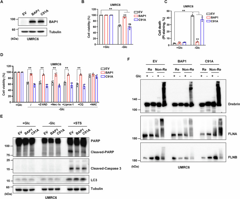
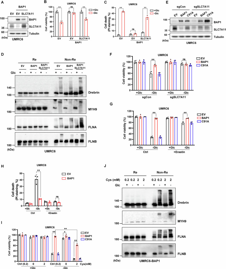
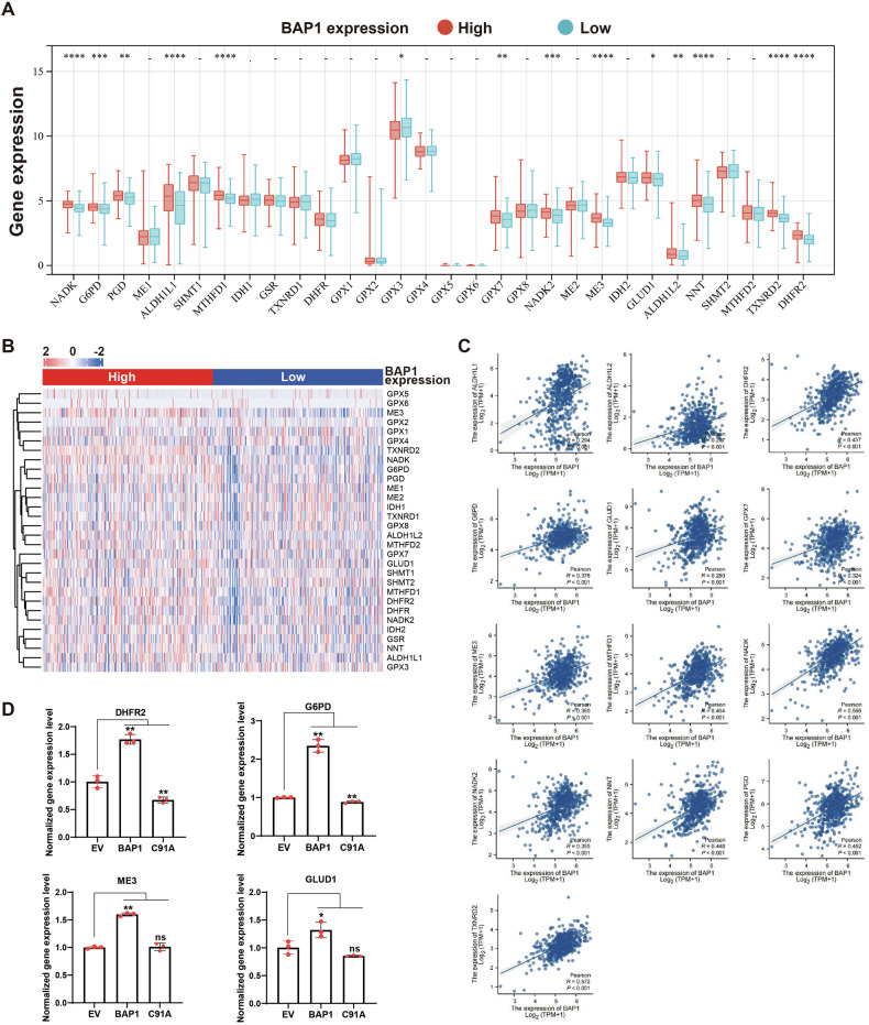
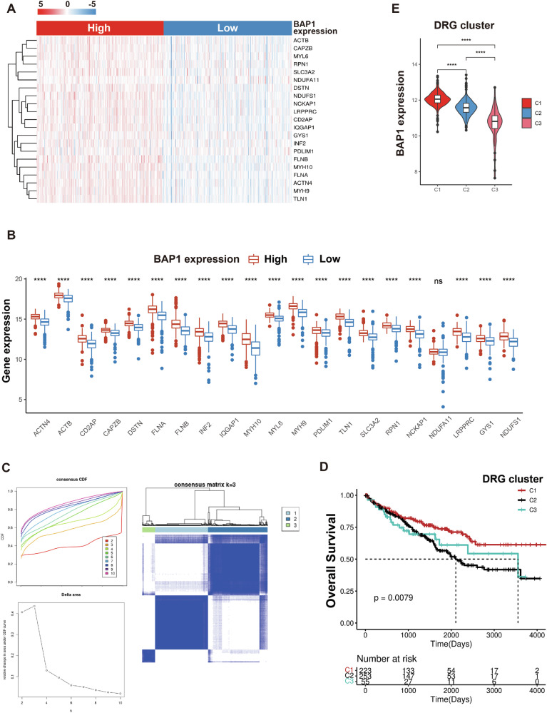
BAP1, BRCA1-Associated Protein 1, serves as a novel tumor suppressor through the deubiquitination of monoubiquitination of H2A and subsequent gene transcriptional regulation. Regulated cell death like apoptosis or ferroptosis is considered an essential mechanism mediating tumor suppression. Previous reports, including ours, have demonstrated that BAP1 could promote apoptosis and ferroptosis to inhibit tumor development. Whether BAP1 regulated additional types of cell death remains unclear. Disulfidptosis is a recently identified novel cell death mode characterized by aberrant accumulation of intracellular disulfide (e.g., cystine) and depletion of NADPH. In this study, we first demonstrated that BAP1 could significantly protect disulfidptosis induced by glucose starvation, which is validated by various cell death inhibitors and the accumulation of disulfide bonds in the cytoskeleton proteins. BAP1 is known to inhibit SLC7A11 expression. We found that the protective effect of BAP1 against disulfidptosis was counteracted when overexpressing SLC7A11 or adding additional cystine. Conversely, BAP1-mediated suppression of disulfidptosis was largely abrogated when SLC7A11-mediated cystine uptake was inhibited by the knockout of SLC7A11 or erastin treatment. Besides, high BAP1 expression showed lower NADP+/NADPH levels, which might confer resistance to disulfidptosis. Consistent with these observations, the expression level of BAP1 was also positively correlated with NADPH-related genes in KIRC patients, though the underlying mechanism mediating NADPH regulation remains further investigation. In summary, our results revealed the role of BAP1 in the regulation disulfidptosis and provided new insights into the understanding of disulfidptosis in tumor development.


Conflict of interest statement

The authors declare no competing interests.

Figures

Fig. 1
Fig. 1. BAP1 suppresses disulfidptosis induced by glucose starvation.
A Validation of the constructed BAP1 WT and BAP1-C91A overexpression in UMRC6 cell lines. B, C Restoring BAP1 WT instead of C91A protects cells from dying upon glucose deprivation. D, E Glucose deprivation induces a unique type of cell death in BAP1-low expression or C91A mutant UMRC6 cells. F Reducing and non-reducing Western blotting analysis of actin cytoskeleton proteins in UMRC6-EV, -BAP1, and -C91A cells upon glucose deprivation condition. The above treatments were terminated when glucose starvation caused significant death of UMRC6-EV cells. Z-VAD, 5 μM; Nec-1s, 2 μM; Liprox-1, 5 μM; CQ, 20 μM; NAC, 2 mM.
Fig. 2
Fig. 2. BAP1 regulates disulfidptosis through the inhibition of SLC7A11-mediated cystine uptake.
A Validation of BAP1 and SLC7A11 dual-overexpressed UMRC6 cell line. B, C SLC7A11 overexpression counteracts the protective role of BAP1 on UMRC6 cells upon glucose deprivation. D SLC7A11 overexpression enhances the actin cytoskeleton proteins tailing under non-reducing conditions in UMRC6-BAP1 cells upon glucose starvation. E Validation of SLC7A11 knockout in UMRC6-EV, -BAP1, and -C91A cells. F Knockout of SLC7A11 saves glucose starvation-induced cell death in UMRC6-EV and -C91A cells. G, H Erastin (10 μM) treatment saves disulfidptosis in UMRC6-EV and C91A cells upon glucose deprivation. I Cystine deprivation saves glucose starvation-induced cell death in UMRC6-EV and -C91A cells, while 2 mM cystine treatment enhances cell death in UMRC6-EV, -BAP1, and -C91A cells. J Cystine at 2 mM aggravated disulfide stress in UMRC6-BAP1 cells cultured in glucose-free and glucose-containing medium. The above treatments were terminated when glucose starvation caused significant death of UMRC6-EV cells.
Fig. 3
Fig. 3. BAP1 decreases NADP+/NADPH ratio within cells.
A Glucose deprivation upregulates NADP+/NADPH ratio in three UMRC6 cell lines (but to a lesser extent in BAP1 cells), and 2DG reverses the upregulation in this case. B 2DG rescues cell death in UMRC6-EV and -C91A cells upon glucose deprivation. C 2DG alleviates the actin cytoskeleton proteins tailing under non-reducing conditions in all three UMRC6 cell lines upon glucose starvation. DG SLC7A11 overexpression (D, F) and 2 mM cystine stimulation (E, G) upregulate NADP+/NADPH ratio and intensify actin cytoskeleton proteins tailing in UMRC6-BAP1 cells upon glucose starvation, which can be rescued by 2DG treatment.
Fig. 4
Fig. 4. Positive correlation between BAP1 and NADPH-related genes expression in KIRC patients.
A Expression level of 30 NADPH-related genes in patients with RCC between BAP1 low and high groups (data from TCGA-KIRC). B Heatmap of the expression levels of NADPH-related genes in BAP1 high and low groups. C Correlation analysis of BAP1 and NADPH-related genes. D Expression levels of significantly differentially expressed NADPH-related genes in UMRC6 cells.
Fig. 5
Fig. 5. The expression level of BAP1 is correlated with disulfidptosis-related genes (DRGs).
A, B Expression level of 21 DRGs in patients with RCC between BAP1 low and high groups (data from TCGA-KIRC). C The CDF distribution diagram and Delta area diagram of consistency clustering analysis shows the optimal κ Value, and the DRG clusters when κ = 3. D The overall survival of patients with RCC varied among different DRG clusters. E BAP1 expression levels in the three DRG clusters.
Fig. 6
Fig. 6. Correlation analysis between BAP1 and DRGs expression in RCC patients with or without cancer-associated BAP1 mutations.
A Expression of BAP1 WT and Mut across different tumor types. B The overall survival of patients with RCC between BAP1 WT and Mut groups. C Expression level of 21 DRGs in patients with RCC between BAP1 WT and Mut groups. D The waterfall diagram shows somatic mutations of BAP1 and DRGs.

References

    1. Jensen DE, Proctor M, Minna J, Borodovsky A, Schultz DC, Wilkinson KD, et al. BAP1: a novel ubiquitin hydrolase which binds to the BRCA1 RING finger and enhances BRCA1-mediated cell growth suppression. Oncogene. 1998;16:1097–112. 10.1038/sj.onc.1201861 - DOI - PubMed
    1. BH Louie, Kurzrock R. BAP1: not just a BRCA1-associated protein. Cancer Treat Rev. 2020;90:102091. 10.1016/j.ctrv.2020.102091 - DOI - PMC - PubMed
    1. Scheuermann JC, de Ayala AA, Okataba K, Ly-Harting N, McGinty RK, Frateman S, et al. Histone H2A deubiquitinase activity of the Polycomb repressive complex PR-DUB. Nature. 2010;465:243–7. 10.1038/nature08966 - DOI - PMC - PubMed
    1. Nishikawa H, Wu W, Koike A, Kojima R, Gomi H, Fukuda M, et al. BRCA1-associated protein 1 interferes with BRCA1/BARD1 RING heterodimer activity. Cancer Res. 2009;69:111–9. 10.1158/0008-5472.CAN-08-3355 - DOI - PubMed
    1. Machida YJ, Machida Y, Vashisht AA, Wohlschlegel JA, Dutta A. The deubiquitinating enzyme BAP1 regulates cell growth via interaction with HCF-1. J Biol Chem. 2009;284:34179–88. 10.1074/jbc.M109.046755 - DOI - PMC - PubMed

LinkOut - more resources