Review
. 2024 Sep 12;8(1):199.
doi: 10.1038/s41698-024-00681-z.

Barriers and opportunities in pancreatic cancer immunotherapy

Affiliations
Review

Barriers and opportunities in pancreatic cancer immunotherapy

Yixin Ju et al. NPJ Precis Oncol. .

Abstract

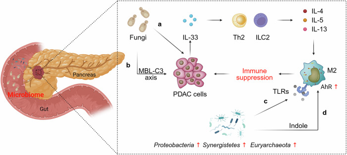
Pancreatic ductal adenocarcinoma (PDAC) presents a fatal clinical challenge characterized by a dismal 5-year overall survival rate, primarily due to the lack of early diagnosis and limited therapeutic efficacy. Immunotherapy, a proven success in multiple cancers, has yet to demonstrate significant benefits in PDAC. Recent studies have revealed the immunosuppressive characteristics of the PDAC tumor microenvironment (TME), including immune cells with suppressive properties, desmoplastic stroma, microbiome influences, and PDAC-specific signaling pathways. In this article, we review recent advances in understanding the immunosuppressive TME of PDAC, TME differences among various mouse models of pancreatic cancer, and the mechanisms underlying resistance to immunotherapeutic interventions. Furthermore, we discuss the potential of targeting cancer cell-intrinsic pathways and TME components to sensitize PDAC to immune therapies, providing insights into strategies and future perspectives to break through the barriers in improving pancreatic cancer treatment.


Conflict of interest statement

The authors declare no competing interests. Figure 2 was created with BioRender.com.

Figures

Fig. 1
Fig. 1. The immunosuppressive TME in PDAC.
a Schematic representation of the interplay among tumor cells, tumor-infiltrating immune cells, and cancer-associated fibroblasts (CAFs) in the PDAC TME. Tumor cells and CAFs secrete chemokines and growth factors, such as granulocyte colony-stimulating factor (G-CSF), granulocyte-macrophage colony-stimulating factor (GM-CSF), CXCL2/5, and CXCL12 to recruit suppressive immune cells to tumor tissues. Pro-tumor immune cells contribute to the exhaustion of effector T cells and the activation of CAFs. Activated CAFs, in turn, support tumor growth through desmoplasia and inflammatory cytokines such as IL-6; in addition, they may cooperate with mast cells to promote tumor cell proliferation and metastasis. Upregulation of immune checkpoint molecules (e.g., PD-L1 and TIGIT) on tumor cells and immune cells, as well as downregulation of MHC-I, contribute to T-cell dysfunction. b Pro-tumor cells include myeloid-derived suppressor cells (MDSCs), M2 macrophages, N2 neutrophils, regulatory T cells, regulatory B cells, mast cells, and Th2 cells. Anti-tumor immune cells include CD8 + T cells, dendritic cells (DC), M1 macrophages, natural killer (NK) cells, N1 neutrophils, and Th1 cells.
Fig. 2
Fig. 2. The microbiome associated with immune suppression in PDAC TME.
a Intratumoral fungus-mediated IL-33 secretion by PDAC cells recruits and activates Th2 cells and innate lymphoid cells 2 (ILC2), which stimulate tumor growth by secreting pro-tumorigenic cytokines such as IL-4, IL-5, and IL-13. b Pathogenic fungi promote PDAC progression by driving the complement cascade via a mannose-binding lectin (MBL)–C3 axis. c Proteobacteria, Synergistetes, and Euryarchaeota are enriched in PDAC patients, reprogramming TAM toward a pro-tumor M2-like phenotype through Toll-like receptors (TLRs). (d) Lactobacillus-derived indole fosters TAM polarization toward an immune-suppressive phenotype through the aryl hydrocarbon receptor (AhR). Figure created with BioRender.com.
Fig. 3
Fig. 3. Tumor-intrinsic signaling pathways associated with immune evasion in PDAC.
a Mutated KRAS (mKRAS), which is permanently bound to GTP, constitutively activates downstream signaling pathways, resulting in PD-L1 overexpression and the recruitment of immunosuppressive cells to pancreatic tumors. b Aberrant activation of the WNT–β-catenin pathway, which can be caused by mutation of RNF43 or lncRNA-mediated inhibition of β-catenin degradation, reduces dendritic cell recruitment by downregulating CCL4 and upregulating PD-L1 expression. c Lipophilic glucocorticoids (GCs) diffuse through the cell membrane and bind to the glucocorticoid receptor (GR) in the cytoplasm of PDAC cells. This binding induces a change in the chaperone complex bound to GR, leading to its translocation into the nucleus. Once in the nucleus, GR activates PD-L1 expression and represses MHC-1 expression by binding to glucocorticoid response elements (GREs), ultimately leading to the reduction in the abundance and effector function of tumor-infiltrating CD8 + T cells.

References

    1. Siegel, R. L., Miller, K. D., Wagle, N. S. & Jemal, A. Cancer statistics, 2023. CA Cancer J. Clin.73, 17–48 (2023). 10.3322/caac.21763 - DOI - PubMed
    1. Park, W., Chawla, A. & O’Reilly, E. M. Pancreatic cancer: a review. JAMA326, 851–862, (2021). 10.1001/jama.2021.13027 - DOI - PMC - PubMed
    1. Mizrahi, J. D., Surana, R., Valle, J. W. & Shroff, R. T. Pancreatic cancer. Lancet395, 2008–2020 (2020). 10.1016/S0140-6736(20)30974-0 - DOI - PubMed
    1. Von Hoff, D. D. et al. Increased survival in pancreatic cancer with nab-paclitaxel plus gemcitabine. N. Engl. J. Med.369, 1691–1703 (2013). 10.1056/NEJMoa1304369 - DOI - PMC - PubMed
    1. Conroy, T. et al. FOLFIRINOX versus gemcitabine for metastatic pancreatic cancer. N. Engl. J. Med. 364, 1817–1825 (2011). 10.1056/NEJMoa1011923 - DOI - PubMed

LinkOut - more resources