. 2024 Oct;31(10):1242-1254.
doi: 10.1038/s41418-024-01370-x. Epub 2024 Sep 12.

Inhibition of nucleo-cytoplasmic proteasome translocation by the aromatic amino acids or silencing Sestrin3-their sensing mediator-is tumor suppressive

Affiliations

Inhibition of nucleo-cytoplasmic proteasome translocation by the aromatic amino acids or silencing Sestrin3-their sensing mediator-is tumor suppressive

Ido Livneh et al. Cell Death Differ. 2024 Oct.

Abstract

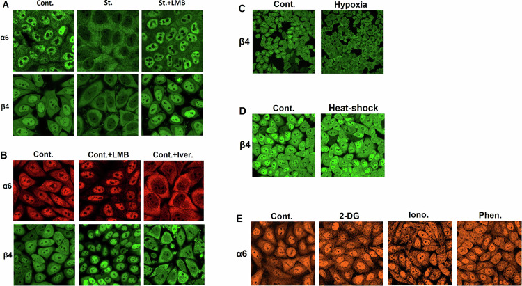
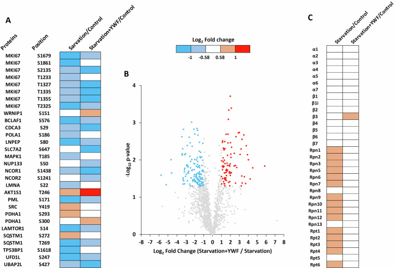
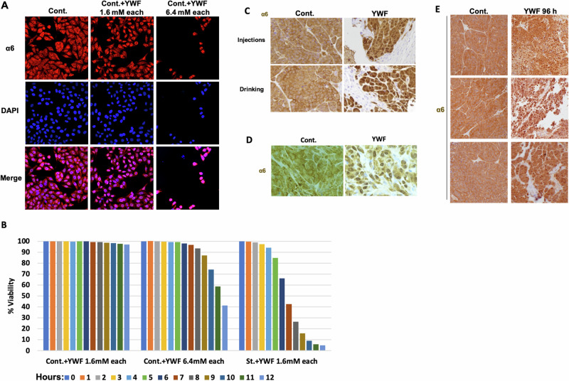
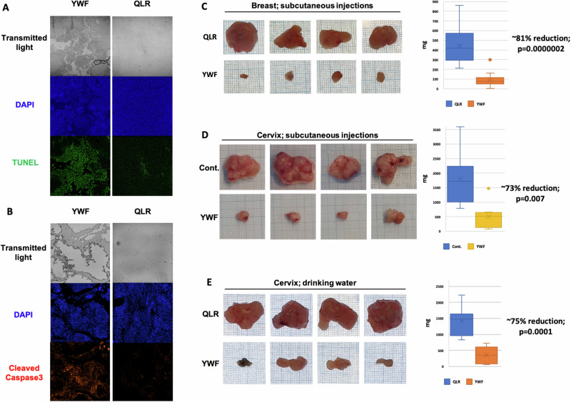
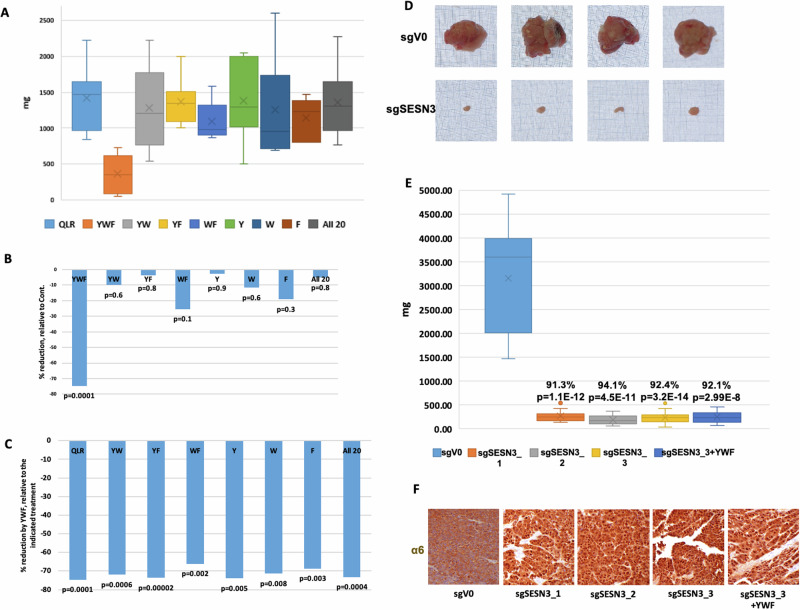
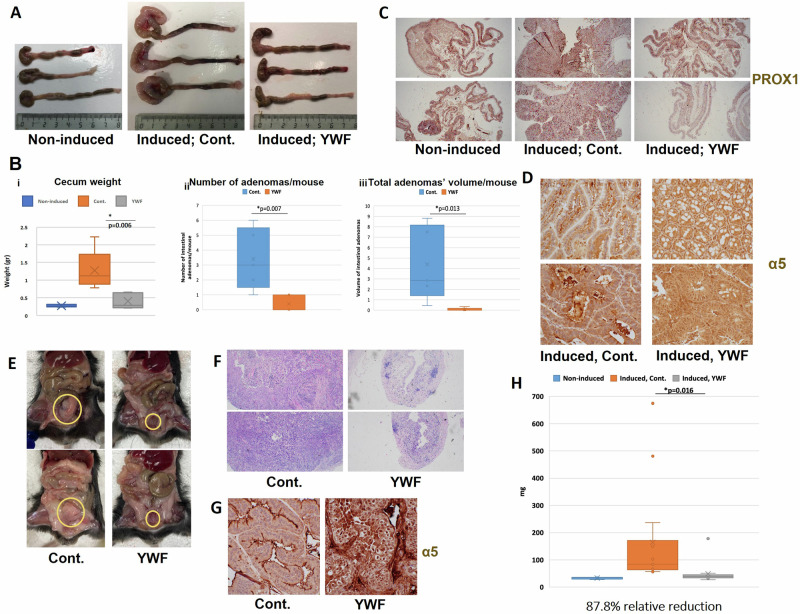
The proteasome, the catalytic arm of the ubiquitin system, is regulated via its dynamic compartmentation between the nucleus and the cytoplasm, among other mechanisms. Under amino acid shortage, the proteolytic complex is translocated to the cytoplasm, where it stimulates proteolysis to supplement recycled amino acids for essential protein synthesis. This response is mediated via the mTOR pathway and the lack of the three aromatic amino acids Tyr, Trp, and Phe (YWF). mTOR activation by supplementation of the triad inhibits proteasome translocation, leading to cell death. We now show that tumoral inherent stress conditions result in translocation of the proteasome from the nucleus to the cytosol. We further show that the modulation of the signaling cascade governed by YWF is applicable also to non-starved cells by using higher concentration of the triad to achieve a surplus relative to all other amino acids. Based on these two phenomena, we found that the modulation of stress signals via the administration of YWF leads to nuclear proteasome sequestration and inhibition of growth of xenograft, spontaneous, and metastatic mouse tumor models. In correlation with the observed effect of YWF on tumors, we found - using transcriptomic and proteomic analyses - that the triad affects various cellular processes related to cell proliferation, migration, and death. In addition, Sestrin3-a mediator of YWF sensing upstream of mTOR-is essential for proteasome translocation, and therefore plays a pro-tumorigenic role, positioning it as a potential oncogene. This newly identified approach for hijacking the cellular "satiety center" carries therefore potential therapeutic implications for cancer.


Conflict of interest statement

IL and AC are co-founders of Tripod Therapeutics Ltd., which develops YWF-based therapies.

Figures

Fig. 1
Fig. 1. Stress-induced translocation of the proteasome from the nucleus to the cytosol is specific but not limited to amino acid shortage.
A HeLa cells were cultured in a complete medium (Cont.) or a starvation medium lacking amino acids in the absence (St.) or presence of Leptomycin B (St.+LMB). The indicated proteasome subunits were visualized via confocal microscopy. B HeLa cells were cultured in a complete medium alone (Cont.), or in the presence of LMB (Cont.+LMB) or ivermectin (Cont.+Iver.), and the proteasome was visualized as above. C HeLa cells were cultured in a complete medium in either 21% (Cont.) or <1% O2 (Hypoxia), and the proteasome was visualized as above. D Hela cells were subjected to heat-shock, and the proteasome was visualized as above. E HeLa cells were treated with either 2-deoxyglucose (2-DG), ionomycin (Iono.), or phenformin (Phen.), and the proteasome was visualized as above.
Fig. 2
Fig. 2. The effect of YWF on gene expression.
HeLa cells were incubated in either a complete medium, starvation medium, starvation medium supplemented with Tyr, Trp, and Phe (St.+YWF), or starvation medium supplemented with Gln, Leu, and Arg (St.+QLR). Cells were harvested, RNA was extracted and sequenced, and differentially expressed genes were compared according to the Ingenuity Pathway Analysis (see “Materials and methods” for further details). The altered functions are clustered according to cell viability/death (A), metabolism (B), and general cell organization and homeostasis (C). Presented are biological functions that were found to be affected differentially in response to the addition of YWF (St.+YWF_vs_Starvation) or QLR (St.+QLR_vs_Starvation).
Fig. 3
Fig. 3. YWF supplementation modulates post-translational modifications and proteasome assembly.
Proteomic experiments were performed to assess changes in protein phosphorylation (A), ubiquitination (B), and proteasome composition (C), following YWF supplementation. Three biological replicates were performed for each experiment, and a two-tailed Welch’s t-test was used. A Differentially phosphorylated proteins are presented in a heat map displaying the Log2 fold-change (Starvation/Control, and Starvation + YWF/Control) for each phosphorylation site. Gene names and phosphorylated residues are presented. B Differentially ubiquitinated proteins are presented in a volcano plot displaying the log2 fold-change (Starvation + YWF/Starvation) for each ubiquitination site quantified, and the corresponding −Log10 p-value. Blue dots represent ubiquitination sites more abundant in starved cells in the absence of YWF; red dots represent those more abundant in the presence of YWF; gray dots represent sites that are not differentially ubiquitinated. C Cell lysates were subjected to immunoprecipitation of the 20S proteasome using an antibody for the α6 subunit. The differentially interacting proteasomal subunits are presented in a heat-map displaying the Log2 fold-change (Starvation/Control, and Starvation + YWF/Control) for each proteasome subunit. The blue, red, and white colors represent a decrease, increase, or no change in the interaction with the α6 subunit, respectively.
Fig. 4
Fig. 4. The YWF-stimulated cascade governs proteasome dynamics also in non-starved cells and animal tumor models.
A HeLa cells were cultured in a complete medium (Cont.), or in the presence of the indicated concentrations of YWF. The proteasome was visualized using confocal microscopy. B HeLa cells were incubated in different YWF concentrations added to a complete DMEM medium in order to assess the cytotoxic effect of the aromatic triad on non-starved cells. Cell viability was quantified using high-throughput fluorescent microscopy. HeLa (C, E) and MDA-MB-231 (D) cells were inoculated in immune-compromised mice to generate xenograft tumors, and animals in the treatment group were administered with YWF (see Materials and methods:lly for the proteasome. Injections refer to the administration of treatment subcutaneously to the tumor bed, while Drinking refers to its administration orally, via the animals’ drinking water.
Fig. 5
Fig. 5. YWF-induced proteasome nuclear sequestration results in cell death and tumor shrinkage.
A HeLa cells were inoculated in immune-compromised mice to generate xenograft tumors, and animals were treated as indicated (see also under “Materials and methods”). Tumors were harvested and tissue sections were fixed in formaldehyde and embedded in paraffin. Presented is the detection of apoptosis using TUNEL staining of histopathological slides. B Histopathological slides were generated as in A, and presented is the detection of apoptosis using staining for cleaved Caspase3. Xenograft tumors were generated as above, using either MDA-MB-231 (C) or HeLa (D) cells. Animals were treated as indicated via subcutaneous injections to the tumor bed. Harvested tumors were weighed and photographed for scale on a graph paper. Plotted are tumor weights at the time of mouse sacrificing. E Xenograft tumors originating from HeLa cells were generated as in (D), and the indicated amino acids were administered via drinking water. Analyses were carried out as in (D).
Fig. 6
Fig. 6. SESN3, mediator of YWF sensing, is a pro-tumorigenic protein.
AC. HeLa cells were inoculated in immune-compromised mice to generate xenograft tumors, and animals were treated as indicated. Tumors were harvested and tissue sections were fixed in formaldehyde and embedded in paraffin. Plotted are tumor weights at the time of mouse sacrificing (A), the reduction in weight following each treatment relative to Control (B), and the reduction in weight by YWF, relative to each of the other indicated combinations of amino acids (C). DF Cells underwent gene editing using CRISPR to generate several clones with knockout of SESN3 (see Livneh et al. [32]). The different clones were inoculated in immune-compromised mice to generate xenograft tumors, and animals were treated as indicated (see also under “Materials and methods”). Tumors were harvested and photographed for scale on a graph paper (D). Presented are tumor weights at the time of mouse sacrificing (E), and immunohistochemical staining of the proteasome in tumor sections (F).
Fig. 7
Fig. 7. Preventing proteasome recruitment inhibits endogenous tumor growth and metastasis.
A Gastrointestinal tumors were induced in immune-competent mice via the loss of the tumor suppressor gene adenomatous polyposis coli (APC; see under “Materials and methods”). Colons and cecums from either non-induced mice, mice in which the loss of the tumor suppressor APC was induced and were either untreated (Cont.) or treated with YWF dissolved in their drinking water. B Tumors generated and treated as in A were harvested, and tissue sections were fixed in formaldehyde and embedded in paraffin. Presented are the plotting of average cecum weight (i), number of tumors along the colon (ii), and their total volume (iii) at the time of mouse sacrificing. C Low magnification of cecums from mice from the indicated groups stained for the high-grade dysplasia marker PROX1. D Immunohistochemistry of the proteasome in histopathological sections from the tumors described under A, B. E The carcinogen BBN was administered to immune-competent mice via their drinking water. Following the formation of urine bladder neoplasms, animals in the treatment group were administered with YWF (see also under “Materials and methods”). Mice were sacrificed, bladders were harvested and tissue sections were fixed in formaldehyde and embedded in paraffin. Presented are gross samples at the time of sacrifice. F Low magnification of H&E staining of bladders from the different experimental groups. G Immunohistochemistry of the proteasome in histopathological section from E. H Plotting of weights of the bladders harvested in E.
Fig. 8
Fig. 8. Nuclear proteasome sequestration using YWF is effective against tumors of different origins, as well as metastasis.
A The carcinogen 3-MCA was injected once into the thigh of immune-competent mice, resulting in the formation of a sarcoma, at which point the animals were treated as indicated (see also under “Materials and methods”). Mice were sacrificed, tumors were harvested, and tissue sections were fixed in formaldehyde and embedded in paraffin. Presented are gross samples from the time of animal sacrifice, photographed for scale on a graph paper. B Immunohistochemistry of the proteasome in histopathological sections from the sarcomas generated as described under A. C Sarcomas were weighed at the time of sacrifice, and tumor weights were plotted, demonstrating the therapeutic effect of YWF. D To generate allograft breast tumors, 4T1 murine breast carcinoma cells expressing a fluorescent marker were inoculated in the mammary glands of female immune-competent mice. Following the formation of tumors, the animals were treated as indicated (see also under “Materials and methods”). Mice were sacrificed, and their livers were isolated and visualized to assess the intensity of fluorescence using IVIS. The fluorescence intensity correlates with the extent of metastases from the primary allograft in the mammary gland. E Analysis of mCherry intensity as measured by IVIS, as described under D.

References

    1. Scheffner M, Nuber U, Huibregtse JM. Protein ubiquitination involving an E1-E2-E3 enzyme ubiquitin thioester cascade. Nature. 1995;373:81–3. - PubMed
    1. Metzger MB, Hristova VA, Weissman AM. HECT and RING finger families of E3 ubiquitin ligases at a glance. J Cell Sci. 2012;125:531–7. - PMC - PubMed
    1. Sun H, Mali SM, Singh SK, Meledin R, Brik A, Kwon YT, et al. Diverse fate of ubiquitin chain moieties: the proximal is degraded with the target, and the distal protects the proximal from removal and recycles. Proc Natl Acad Sci USA. 2019;116:7805–12. - PMC - PubMed
    1. Gomez TA, Kolawa N, Gee M, Sweredoski MJ, Deshaies RJ. Identification of a functional docking site in the Rpn1 LRR domain for the UBA-UBL domain protein Ddi1. BMC Biol. 2011;9:33. - PMC - PubMed
    1. Zheng N, Shabek N. Ubiquitin ligases: structure, function, and regulation. 2017. 10.1146/annurev-biochem - PubMed

LinkOut - more resources