

Automated Three-Dimensional Imaging and Pfirrmann Classification of Intervertebral Disc Using a Graphical Neural Network in Sagittal Magnetic Resonance Imaging of the Lumbar Spine
- PMID: 39266913
- PMCID: PMC11950579
- DOI: 10.1007/s10278-024-01251-2
Automated Three-Dimensional Imaging and Pfirrmann Classification of Intervertebral Disc Using a Graphical Neural Network in Sagittal Magnetic Resonance Imaging of the Lumbar Spine
Abstract
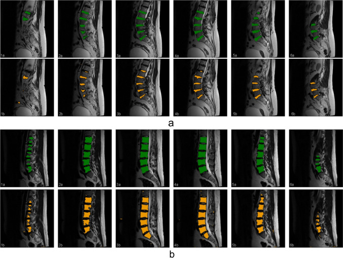
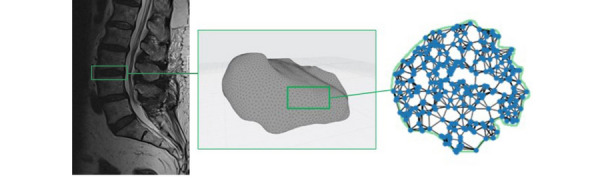
This study aimed to develop a graph neural network (GNN) for automated three-dimensional (3D) magnetic resonance imaging (MRI) visualization and Pfirrmann grading of intervertebral discs (IVDs), and benchmark it against manual classifications. Lumbar IVD MRI data from 300 patients were retrospectively analyzed. Two clinicians assessed the manual segmentation and grading for inter-rater reliability using Cohen's kappa. The IVDs were then processed and classified using an automated convolutional neural network (CNN)-GNN pipeline, and their performance was evaluated using F1 scores. Manual Pfirrmann grading exhibited moderate agreement (κ = 0.455-0.565) among the clinicians, with higher exact match frequencies at lower lumbar levels. Single-grade discrepancies were prevalent except at L5/S1. Automated segmentation of IVDs using a pretrained U-Net model achieved an F1 score of 0.85, with a precision and recall of 0.83 and 0.88, respectively. Following 3D reconstruction of the automatically segmented IVD into a 3D point-cloud representation of the target intervertebral disc, the GNN model demonstrated moderate performance in Pfirrmann classification. The highest precision (0.81) and F1 score (0.71) were observed at L2/3, whereas the overall metrics indicated moderate performance (precision: 0.46, recall: 0.47, and F1 score: 0.46), with variability across spinal levels. The integration of CNN and GNN offers a new perspective for automating IVD analysis in MRI. Although the current performance highlights the need for further refinement, the moderate accuracy of the model, combined with its 3D visualization capabilities, establishes a promising foundation for more advanced grading systems.
Keywords: Artificial intelligence; Convolutional neural network; Graph neural network; Intervertebral disc degeneration; MRI; Machine learning; Pfirrmann classification; Spine imaging.
© 2024. The Author(s).
Conflict of interest statement
Declarations. Ethics Approval: This study protocol was approved by the ethics committee of the Medical Faculty at the University of Leipzig, Germany (Ethics Committee; 025/21-ek) and in accordance with the 1964 Helsinki Declaration and its later amendments or comparable ethical standards. Consent to Participate: Informed consent was obtained from all individual participants included in the study. Competing Interests: The authors declare no competing interests.
Figures
Similar articles
-
Novel Application of the Pfirrmann Disc Degeneration Grading System to 9.4T MRI: Higher Reliability Compared to 3T MRI.Spine (Phila Pa 1976). 2019 Jul 1;44(13):E766-E773. doi: 10.1097/BRS.0000000000002967. Spine (Phila Pa 1976). 2019. PMID: 31205169
-
The importance of level stratification for quantitative MR studies of lumbar intervertebral discs: a cross-sectional analysis in 101 healthy adults.Eur Spine J. 2019 Sep;28(9):2153-2161. doi: 10.1007/s00586-019-06059-1. Epub 2019 Jul 12. Eur Spine J. 2019. PMID: 31309335
-
Lumbar spine MRI annotation with intervertebral disc height and Pfirrmann grade predictions.PLoS One. 2024 May 10;19(5):e0302067. doi: 10.1371/journal.pone.0302067. eCollection 2024. PLoS One. 2024. PMID: 38728318 Free PMC article.
-
Measurement of Three-Dimensional Internal Dynamic Strains in the Intervertebral Disc of the Lumbar Spine With Mechanical Loading and Golden-Angle Radial Sparse Parallel-Magnetic Resonance Imaging.J Magn Reson Imaging. 2021 Aug;54(2):486-496. doi: 10.1002/jmri.27591. Epub 2021 Mar 13. J Magn Reson Imaging. 2021. PMID: 33713520 Free PMC article. Review.
-
Association between non-traumatic vertebral fractures and adjacent discs degeneration: a cross-sectional study and literature review.BMC Musculoskelet Disord. 2020 Nov 27;21(1):781. doi: 10.1186/s12891-020-03814-0. BMC Musculoskelet Disord. 2020. PMID: 33246433 Free PMC article. Review.
Cited by
-
A CNN Autoencoder for Learning Latent Disc Geometry from Segmented Lumbar Spine MRI.medRxiv [Preprint]. 2025 Mar 4:2025.02.28.25323111. doi: 10.1101/2025.02.28.25323111. medRxiv. 2025. PMID: 40093260 Free PMC article. Preprint.
-
Segment Anything Model (SAM) and Medical SAM (MedSAM) for Lumbar Spine MRI.Sensors (Basel). 2025 Jun 7;25(12):3596. doi: 10.3390/s25123596. Sensors (Basel). 2025. PMID: 40573483 Free PMC article.
References
-
- Pfirrmann CW, Metzdorf A, Zanetti M, Hodler J, Boos N. Magnetic resonance classification of lumbar intervertebral disc degeneration. Spine 2001; 26(17):1873–8. - PubMed
-
- Arana E, Royuela A, Kovacs FM, Estremera A, Sarasíbar H, Amengual G et al. Lumbar spine: agreement in the interpretation of 1.5-T MR images by using the Nordic Modic Consensus Group classification form. Radiology 2010; 254(3):809–17. - PubMed
MeSH terms
Grants and funding
LinkOut - more resources
Full Text Sources
Medical