. 2024 Sep 12;15(1):298.
doi: 10.1186/s13287-024-03919-6.

Expandable hESC-derived cardiovascular progenitor cells generate functional cardiac lineage cells for microtissue construction

Affiliations

Expandable hESC-derived cardiovascular progenitor cells generate functional cardiac lineage cells for microtissue construction

Siamak Rezaeiani et al. Stem Cell Res Ther. .

Abstract

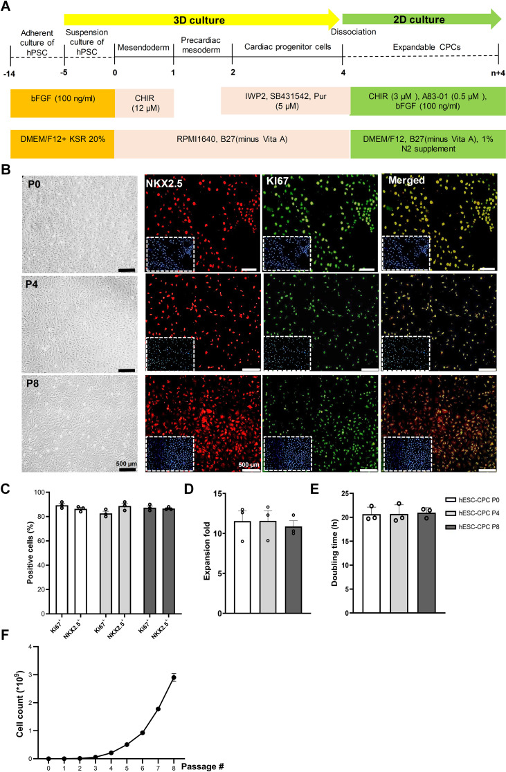
Background: Cardiovascular progenitor cells (CPCs) derived from human embryonic stem cells (hESCs) are considered valuable cell sources for investigating cardiovascular physiology in vitro. Meeting the diverse needs of this application requires the large-scale production of CPCs in an in vitro environment. This study aimed to use an effective culture system utilizing signaling factors for the large-scale expansion of hESC-derived CPCs with the potential to differentiate into functional cardiac lineage cells.

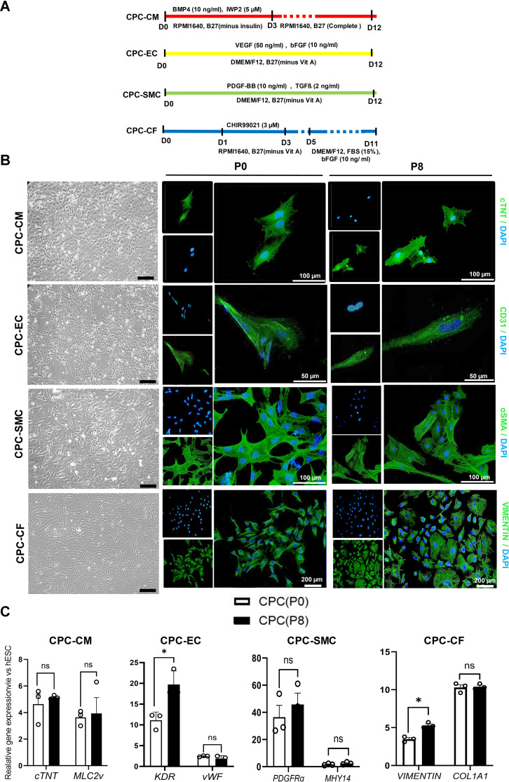
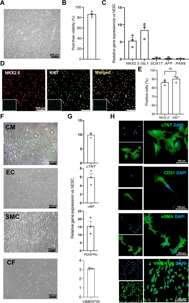
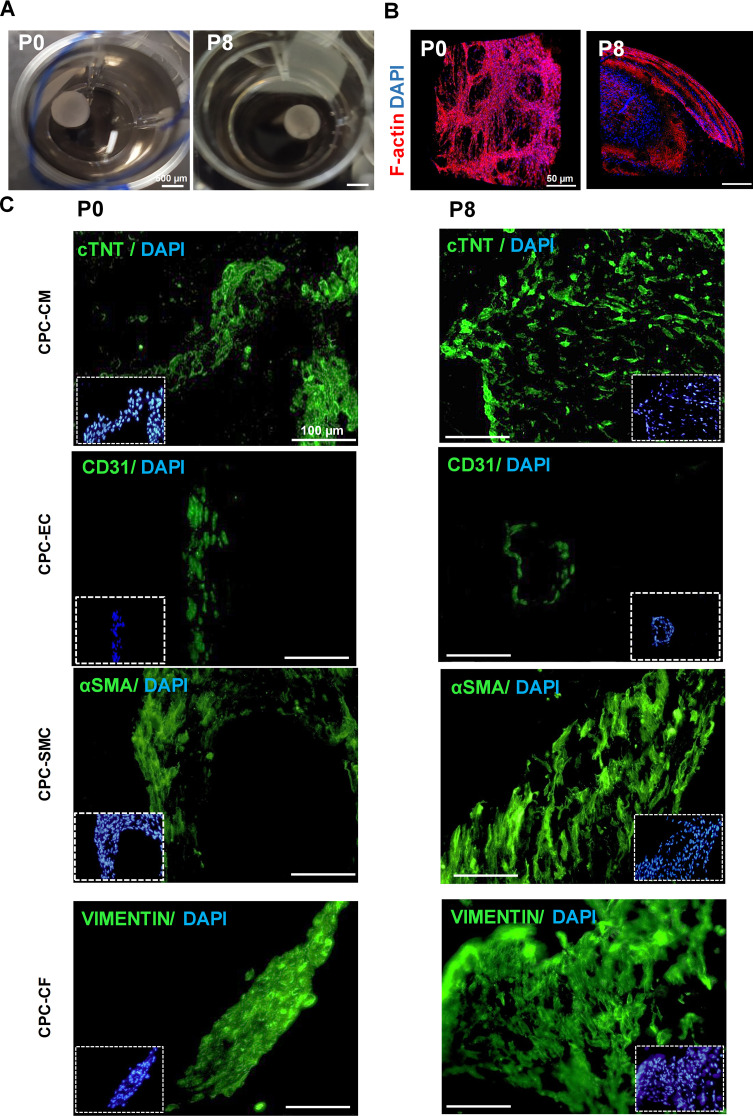
Methods and results: Initially, CPCs were generated from hESCs using a 4-day differentiation protocol with a combination of four small molecules (CHIR99021, IWP2, SB-431542, and purmorphamine). These CPCs were then expanded and maintained in a medium containing three factors (bFGF, CHIR, and A83-01), resulting in a > 6,000-fold increase after 8 passages. These CPCs were successfully cryopreserved for an extended period in late passages. The expanded CPCs maintained their gene and protein expression signatures as well as their differentiation capacity through eight passages. Additionally, these CPCs could differentiate into four types of cardiac lineage cells: cardiomyocytes, endothelial cells, smooth muscle cells, and fibroblasts, demonstrating appropriate functionality. Furthermore, the coculture of these CPC-derived cardiovascular lineage cells in rat tail collagen resulted in cardiac microtissue formation, highlighting the potential of this 3D platform for studying cardiovascular physiology in vitro.

Conclusion: In conclusion, expandable hESC-derived CPCs demonstrated the ability to self-renewal and differentiation into functional cardiovascular lineage cells consistently across passages, which may apply as potential cell sources for in vitro cardiovascular studies.

Keywords: Cardiovascular progenitor cells; Cryopreservation; Differentiation; Expansion; Large-scale production.


Conflict of interest statement

The authors declare that they have no competing interests.

Figures

Fig. 1
Fig. 1
Characterization of cardiac progenitor cells (CPCs) generated from the human embryonic stem cell (hESC). (A) Schematic diagram showing differentiation protocol used for CPC induction from hESCs. hESCs were cultured in suspension as spheroids and differentiated into mesoderm, followed by cardiac progenitor cells differentiation by one-day treatment with 12 µM CHIR99021 (CHIR) and a one-day rest period, then treatment with ISP 5 µM for two-days. The 4-day old differentiated hESC-CPC spheroids were dissociated into single cells and cultured in expansion medium. (B) CPC morphology and immunofluorescence staining of NKX2.5 and Ki67 expressions in P0, 4, 8 CPCs cultured in expansion medium. Cells were counter-stained with DAPI (C) Numeric data of percentages of NKX2.5+ and KI67+ cells in hESC-derived CPCs. Data are presented as mean ± standard error of the mean (SEM). (D & E) Doubling time and expansion fold of CPCs at 0, 4, 8 passages. Data are presented as mean ± standard error of the mean (SEM). (F) Representative graph of total cell counts generated after 8 passages of hESC-derived CPCs
Fig. 2
Fig. 2
Differentiation and characterization of cardiomyocyte (CM), endothelial cell (EC), smooth muscle cell (SMC), and cardiac fibroblasts (CF) from CPCs. (A) Schematic diagram of differentiation protocol of CM, EC, SMC, and CF. (B) Morphology and immunofluorescence characterization of CM (with c-TNT), EC (with CD31), SMC (with αSMA), and CF (with VIMENTIN) derived from CPCs at both passages 0 and 8. Scale bar; 200 μm. (C) Characterization of four cardiac lineage cells derived from CPCs at both passages 0 and 8 at gene expression level: c-TNT and MLC2v for CM, KDR and vWF for EC, PDGFRα and MHY14 for SMC, and VIMENTIN and COL1A1 for CF. Data are presented as mean ± standard error of the mean (SEM)
Fig. 3
Fig. 3
Functional assays for cardiac linaege cells derived from both passages 0 and 8. (A & B) The CPC-derived CMs demonstrated regular field potentials. (C) Tube formation was evaluated in CPC-derived ECs and human umbilical vein endothelial cells (HUVEC) within 2 h. (D) Numerical measurements of number of the branches, nodes, and junctions’ formation of CPC-derived ECs, which was compared with HUVEC, as a control group, representing the comparable ability of tube formation of CPC-ECs. (E) Contractility assay of CPC-derived SMCs using carbachol 10 µM during 10 min. Shortening of SMCs were obviously seen, indicating the ability of contraction of these cells. (F) Numerical assessment also represented a significant reduction in cell area. (G & H) Expression of αSMA in activated CFs with doxorubicin was found at protein level (immunostaining) as well as gene expression level (qRT-PCR). All data are presented as mean ± standard error of the mean (SEM)
Fig. 4
Fig. 4
Assessment of the cryopreservation of CPCs. (A) Morphology of CPCs after freeze-thaw. (B) Assessing the percentage of viable cells after freeze-thaw that showed more than 80% viablility. (C) Comparison of the gene expression of freeze-thawed CPCs and hESCs. The upregulation of NKX2.5 and ISL1 and downregulation of SOX17, AFP, and PAX6 were observed copmared to hESCs, reflecting the preservation of CPCs transcriptional signature after freeze-thaw. (D & E) Assessing the NKX2.5 and Ki67 positive CPCs in immunofluorescence, showing more than 80% and 90% of cells were positive for NKX2.5 and Ki67, respectively. (F & G & H) Evaluation of morphology, gene expression, and immunofluorescence staining of the CMs, ECs, SMCs, and CFs differentiated from CPCs after freeze-thaw, implying cryopreservation of differentiation capacity of the CPCs. All data are presented as mean ± standard error of the mean (SEM)
Fig. 5
Fig. 5
Microtissue formation using CM, EC, SMC, and CF in ratio of 2:1:1:2 in collagen derived from rat tail. (A) Macroscopic morphology of the microtissue. (B) Cellularity of the microtissue identified using Rhodamine/Phalloidin staining to show F-actin distribution in the cells, exhibited with Confocal microscope. (C). Detection of CMs, ECs, SMCs, and CFs in the microtissue using immunofluorescence staining: CMs with c-TNT, ECs with CD31, SMCs with αSMA, and CFs with Vimentin

References

    1. Roth GA, Mensah GA, Johnson CO, Addolorato G, Ammirati E, Baddour LM et al. Global burden of cardiovascular diseases and risk factors, 1990–2019: update from the GBD 2019 study. 2020;76(25):2982–3021. - PMC - PubMed
    1. Leone M, Magadum A, Engel FB. Cardiomyocyte proliferation in cardiac development and regeneration: a guide to methodologies and interpretations. Am J Physiol Heart Circ Physiol. 2015;309(8):H1237–50. 10.1152/ajpheart.00559.2015 - DOI - PubMed
    1. Guo Y, Yu Y, Hu S, Chen Y, Shen ZJCD, Disease. Therapeutic Potential Mesenchymal stem Cells Cardiovasc Dis. 2020;11(5):349. - PMC - PubMed
    1. Barreto S, Hamel L, Schiatti T, Yang Y, George VJC. Cardiac progenitor cells from stem cells: learning from genetics and biomaterials. 2019;8(12):1536. - PMC - PubMed
    1. Burridge PW, Keller G, Gold JD, Wu JCJC. Production of de novo cardiomyocytes: human pluripotent stem cell differentiation and direct reprogramming. 2012;10(1):16–28. - PMC - PubMed

LinkOut - more resources