. 2024 Aug 29:37:13232.
doi: 10.3389/ti.2024.13232. eCollection 2024.

Antibody-Mediated Rejection in Liver Transplantation: Immuno-Pathological Characteristics and Long-Term Follow-Up

Affiliations

Antibody-Mediated Rejection in Liver Transplantation: Immuno-Pathological Characteristics and Long-Term Follow-Up

Luca Cicalese et al. Transpl Int. .

Abstract

The diagnosis of liver antibody-mediated rejection (AMR) is challenging and likely under-recognized. The association of AMR with donor-specific antibodies (DSA), and its clinical course in relation to pathologic findings and treatment are ill defined. We identified cases of liver AMR by following the criteria outlined by the 2016 Banff Working Group. Patient demographics, native liver disease, histopathologic findings, treatment type, clinical outcome, and transaminase levels during AMR diagnosis, treatment, and resolution were determined. Patients (n = 8) with AMR average age was 55.2 years (range: 19-68). Seven of eight cases met the Banff criteria for AMR. Personalized treatment regimens consisted of optimization of immunosuppression, intravenous pulse steroids, plasmapheresis, IVIG, rituximab, and bortezomib. Five patients experienced complete resolution of AMR, return of transaminases to baseline, and decreased DSA at long-term follow-up. One patient developed chronic AMR and two patients required re-transplantation. Follow-up after AMR diagnosis ranged from one to 11 years. Because AMR can present at any time, crossmatch, early biopsy, and routine monitoring of DSA levels should be implemented following transaminase elevation to recognize AMR. Furthermore, treatment should be immediately implemented to reverse AMR and prevent graft failure, chronic damage, re-transplantation, and possibly mortality.

Keywords: AMR; C4d; DSA; allograft; rejection; solid organ transplant.


Conflict of interest statement

The authors declare that the research was conducted in the absence of any commercial or financial relationships that could be construed as a potential conflict of interest.

Figures
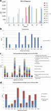
FIGURE 1
FIGURE 1
(A) Serum DSA levels at the time of diagnosis of AMR in the eight patients. (B) h-score of eight AMR patients according to 2016 Banff Criteria for liver allografts. Of these, seven out of eight patients are greater than one. Patient #2 showed minimal pathologic changes in liver biopsy and h-score is zero. (C) Semi-quantitative histology scores of acute AMR in patients where “C” denotes matched control patients. (D) Semi-quantitative histology scores of chronic AMR in patients “C” denotes matched control patients.
FIGURE 2
FIGURE 2
Representative liver biopsy histology of patient #3 at diagnosis and 6-month follow-up. (A–D) Liver biopsy of the patient at the time of diagnosis showed the classic acute AMR microvascular pathology lesions. (E–H) Liver biopsy of the same patient at time of 6-month follow-up.
FIGURE 3
FIGURE 3
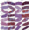
Representative histology of AMR patients at the time of diagnosis (first two columns) and 6–12 months follow-up (last two columns). Each biopsy shows H&E and Masson’s trichrome stain (fibrotic areas are blue). Black scale is 200 µm. (A1–A4) are the biopsies from AMR patient #1. (B1–B4) are the biopsies from AMR patient #2. (C1–C4) are the biopsies from AMR patient #3. (D1–D4) are the biopsies from AMR patient #4. (E1–E4) are the biopsies from AMR patient #5. (F1–F4) are the biopsies from AMR patient #6. (G1–G4) are the biopsies from AMR patient #8. Patient #7 did not have a follow up biopsy after diagnosis.
FIGURE 4
FIGURE 4
Representative liver-related laboratory testing of total bilirubin and the enzymes alanine transaminase (ALT), aspartate aminotransferase (AST), and alkaline phosphatase (ALP) in patient #3. Arrow indicates the time point at the start of treatment.

References

    1. Demetris AJ, Jaffe R, Tzakis A, Ramsey G, Todo S, Belle S, et al. Antibody-Mediated Rejection of Human Orthotopic Liver Allografts. A Study of Liver Transplantation Across ABO Blood Group Barriers. Am J Pathol (1988) 132(3):489–502. - PMC - PubMed
    1. Krukemeyer MG, Moeller J, Morawietz L, Rudolph B, Neumann U, Theruvath T, et al. Description of B Lymphocytes and Plasma Cells, Complement, and Chemokines/Receptors in Acute Liver Allograft Rejection. Transplantation (2004) 78(1):65–70. 10.1097/01.TP.0000132324.14207.8B - DOI - PubMed
    1. Ge X, Ericzon BG, Nowak G, Ohrstrom H, Broom U, Sumitran-Holgersson S. Are Preformed Antibodies to Biliary Epithelial Cells of Clinical Importance in Liver Transplantation? Liver Transplant (2003) 9(11):1191–8. 10.1053/jlts.2003.50236 - DOI - PubMed
    1. Muro M, Legaz I. Importance of Human Leukocyte Antigen Antibodies and Leukocyte Antigen/Killer-Cell Immunoglobulin-Like Receptor Genes in Liver Transplantation. World J Gastroenterol (2023) 29(5):766–72. 10.3748/wjg.v29.i5.766 - DOI - PMC - PubMed
    1. Demetris A, Murase N, Nakamura K, Iwaki Y, Yagihashi A, Valdivia L, et al. Immunopathology of Antibodies as Effectors of Orthotopic Liver Allograft Rejection. Semin Liver Dis (1992) 12(01):51–9. 10.1055/s-2007-1007376 - DOI - PubMed

LinkOut - more resources