Review
. 2024 Aug 29:15:1465775.
doi: 10.3389/fimmu.2024.1465775. eCollection 2024.

Neuroendocrine neoplasms of the thymus

Affiliations
Review

Neuroendocrine neoplasms of the thymus

Paul D Barone et al. Front Immunol. .

Abstract

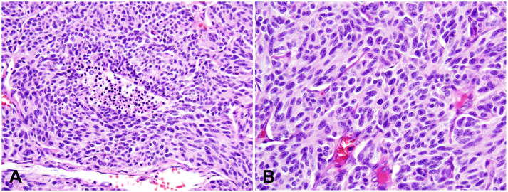
Neuroendocrine neoplasms of the thymus (tNENs), including typical carcinoid, atypical carcinoid, large cell neuroendocrine carcinoma, and small cell carcinoma, are rare tumors with scarce clinical and pathological data available in the literature. They share many common features with neuroendocrine neoplasms in other organs, such as those in the lungs, while demonstrating some distinct clinical and pathological features. This review aims to give an updated overview of each category of tNENs, focusing primarily on the pathologic diagnosis and differential diagnosis of these tumors.

Keywords: carcinoid; large cell neuroendocrine carcinoma; neuroendocrine neoplasm; small cell carcinoma; thymus.


Conflict of interest statement

The authors declare that the research was conducted in the absence of any commercial or financial relationships that could be construed as a potential conflict of interest.

Figures

Figure 1
Figure 1
Typical carcinoid (TC). (A-D) Low to intermediate magnification photomicrographs of TCs show that tumor cells are arranged in trabeculae (A), cords and ribbons (B), glandular structures (C), or solid nests (D), with fine fibrous septa and rich, delicate vasculatures. Magnification (A-D): 40X. (E) High magnification view of TC comprising bland tumor cells with a moderate amount of pale eosinophilic cytoplasm, round to oval nuclei, and finely granular, salt-and-pepper chromatin. The cells are arranged in organoid solid nests separated with delicate vascular stroma. There is no mitosis or necrosis. Magnification (E): 400X. H&E stain (A-E). (F) The Ki-67 index is low (<2%).
Figure 2
Figure 2
Atypical carcinoid (AC). (A) Intermediate magnification photomicrograph of AC with a small focus of comedo-type necrosis within the tumor nest. Magnification: 100X. (B) High magnification view of AC with a mitotic figure in the center of this photo. Tumor cells show neuroendocrine cytologic features, including a moderate amount of eosinophilic cytoplasm and finely granular nuclear chromatin. Magnification: 400X. H&E stain (A, B).
Figure 3
Figure 3
Large cell neuroendocrine carcinoma (LCNEC). (A) High magnification view of LCNEC comprising large pleomorphic tumor cells with a moderate amount of eosinophilic cytoplasm, coarse chromatin, and prominent nucleoli. Frequent mitoses and apoptotic bodies are seen. The tumor shows a nested growth pattern with focal peripheral palisading. H&E stain. (B) Immunohistochemical stain of Chromogranin A is diffusely positive in the tumor cells. Magnification (A, B): 400X. Photographs are courtesy of Jeffrey L. Myers, MD, University of Michigan, Ann Arbor, MI.
Figure 4
Figure 4
Small cell carcinoma (SmCC). (A) High magnification view of SmCC showing densely packed small to intermediate-sized tumor cells with scant cytoplasm, dense chromatin, and occasional inconspicuous nucleoli. Numerous mitoses and apoptotic bodies are seen. (B) One area of the tumor shows a prominent crushing artifact, a common feature of SmCC. H&E stain (A, B). (C) The tumor cells show characteristic cytoplasmic perinuclear dot-like staining for cytokeratin Cam 5.2. (D) The Ki-67 index is high (almost 100%). Magnification (A-D): 400X.

References

    1. Moran CA, Suster S. Neuroendocrine carcinomas (carcinoid tumor) of the thymus. A clinicopathologic analysis of 80 cases. Am J Clin Pathol. (2000) 114:100–10. doi: 10.1309/3PDN-PMT5-EQTM-H0CD - DOI - PubMed
    1. Goto K, Kodama T, Matsuno Y, Yokose T, Asamura H, Kamiya N, et al. . Clinicopathologic and DNA cytometric analysis of carcinoid tumors of the thymus. Mod Pathol. (2001) 14:985–94. doi: 10.1038/modpathol.3880423 - DOI - PubMed
    1. Bohnenberger H, Dinter H, Konig A, Strobel P. Neuroendocrine tumors of the thymus and mediastinum. J Thorac Dis. (2017) 9:S1448–57. doi: 10.21037/jtd.2017.02.02 - DOI - PMC - PubMed
    1. Dinter H, Bohnenberger H, Beck J, Bornemann-Kolatzki K, Schutz E, Kuffer S, et al. . Molecular classification of neuroendocrine tumors of the thymus. J Thorac Oncol. (2019) 14:1472–83. doi: 10.1016/j.jtho.2019.04.015 - DOI - PubMed
    1. Ströbel AMM P, Marom EM, Pelosi G. Thymic neuroendocrine neoplasms. In: WHO Classification of Tumours: Thoracic tumours, 5th ed. Lyon, France: International Agency for Research on Cancer; (2021). Board WCoTE, ed.

Substances

LinkOut - more resources