

Spatial memory and learning: investigating the role of dynamic visual acuity
- PMID: 39267984
- PMCID: PMC11390580
- DOI: 10.3389/fnbeh.2024.1429069
Spatial memory and learning: investigating the role of dynamic visual acuity
Abstract
Introduction: The vestibular system's contribution to spatial learning and memory abilities may be clarified using the virtual Morris Water Maze Task (vMWMT). This is important because of the connections between the vestibular system and the hippocampus area. However, there is ongoing debate over the role of the vestibular system in developing spatial abilities. This study aimed to evaluate the relationship between Dynamic Visual Acuity (DVA) across three planes and spatial abilities.
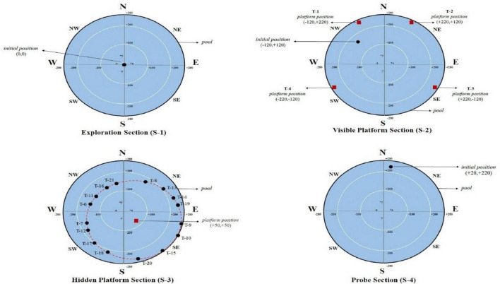
Methods: This cross-sectional study was conducted with 50 healthy adults aged 18 to 55 with normal stress levels and mental health and no neurological, audiological, or vestibular complaints. The Trail-Making Test (TMT) Forms A and B for the assessment of executive functions, the DVA test battery for the evaluation of visual motor functions, and the Virtual Morris Water Maze Test (vMWMT) for the assessment of spatial learning and spatial memory were performed. All participants also underwent the Benton Face Recognition Test (BFRT) and Digit Symbol Substitution Tests (DSST) to assess their relation with spatial memory.
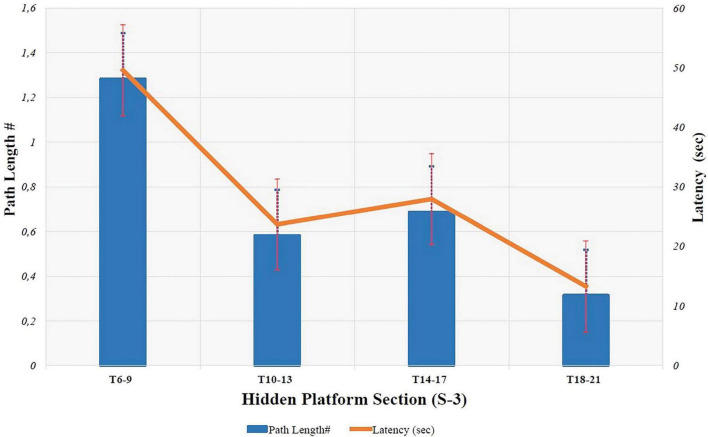
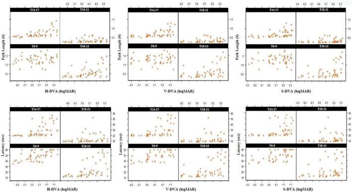
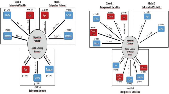
Results: DVA values in horizontal (H-DVA), vertical (V-DVA), and sagittal (S-DVA) planes ranged from (-0.26) to 0.36 logMAR, (-0.20) to 0.36 logMAR, and (-0.28) to 0.33 logMAR, respectively. The latency of three planes of DVA was affected by vMWMT (Horizontal, Vertical, and Sagittal; Estimate: 22.733, 18.787, 13.341, respectively p < 0.001). Moreover, a moderately significant correlation was also found, with a value of 0.571 between the Virtual MWM test and BFRT and a value of 0.539 between the DSST (p < 0.001).
Conclusion: Spatial abilities in healthy adults were significantly influenced by dynamic visual functions across horizontal, vertical, and sagittal planes. These findings are expected to trigger essential discussions about the mechanisms that connect the vestibular-visual system to the hippocampus. The original vMWMT protocol is likely to serve as a model for future studies utilizing this technology.
Keywords: dynamic visual acuity; spatial learning; spatial memory; trail-making test; virtual Morris Water Maze Test.
Copyright © 2024 Kabiş, Gürses, Işıkay and Aksoy.
Conflict of interest statement
The authors declare that the research was conducted in the absence of any commercial or financial relationships that could be construed as a potential conflict of interest.
Figures
References
-
- Barmack N. H., Yakhnitsa V. (2021). “Vestibulocerebellar functional connections,” in Handbook of the cerebellum and cerebellar disorders, eds Manto M. U., Gruol D. L., Schmahmann J. D., Koibuchi N., Sillitoe R. V. (Cham: Springer; ), 467–495.
-
- Benton A., Van Allen M. (1968). Impairment in facial recognition in patients with cerebral disease. Cortex 4 344–IN1. - PubMed
LinkOut - more resources
Full Text Sources