. 2024 Jun 17;8(5):102475.
doi: 10.1016/j.rpth.2024.102475. eCollection 2024 Jul.

Modeling cancer-associated hypercoagulability using glioblastoma spheroids in microfluidic chips

Affiliations

Modeling cancer-associated hypercoagulability using glioblastoma spheroids in microfluidic chips

Maaike Y Kapteijn et al. Res Pract Thromb Haemost. .

Abstract

Background: Cancer increases the risk of venous thromboembolism, and glioblastoma is one of the cancer types with the highest risk of venous thromboembolism (10%-30%). Tumor-intrinsic features are believed to affect vascular permeability and hypercoagulability, but novel models are required to study the pathophysiological dynamics underlying cancer-associated thrombosis at the molecular level.

Objectives: We have developed a novel cancer-on-a-chip model to examine the effects of glioblastoma cells on the deregulation of blood coagulation.

Methods: This was accomplished by coculturing vessel-forming human umbilical vein endothelial cells with glioblastoma spheroids overexpressing tissue factor (TF), the initiator of coagulation (U251 lentivirus, LV-TF) or an LV-control (U251 LV-Ctrl) in an OrganoPlate Graft platform.

Results: Using a modified thrombin generation assay inside the cancer-on-a-chip, we found that U251 LV-Ctrl and U251 LV-TF spheroids promoted an increased procoagulant state in plasma, as was shown by a 3.1- and 7.0-fold increase in endogenous thrombin potential, respectively. Furthermore, the anticoagulant drug rivaroxaban and TF coagulation-blocking antibody 5G9 inhibited the activation of blood coagulation in U251 LV-TF spheroid-containing graft plates, as was shown by a reduced endogenous thrombin potential (4.0- and 4.4-fold, respectively).

Conclusion: With this study, we present a novel 3-dimensional cancer-on-a-chip model that has the potential to be used in the discovery of new anticoagulant drugs and identification of optimal anticoagulant strategies for glioblastoma and other cancer types.

Keywords: anticoagulants; cancer-associated thrombosis; extracellular vesicles; glioblastoma; organ-on-a-chip.


Figures

Figure 1
Figure 1
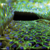
Cancer-associated hypercoagulability-on-a-chip for glioblastoma. (A) Bottom view of an OrganoPlate Graft consisting of 64 independent perfusable tissue culture chips. (B) Schematic upper view representation of one OrganoPlate Graft tissue culture chip. (C) Side view of a chip showing growth of human umbilical vein endothelial cell (HUVEC) vessels in cell culture medium next to the extracellular matrix, consisting of collagen type I. The white arrows represent the bidirectional flow. (D) Schematic depiction of the experimental setup. (E) Example of a live coculture of HUVEC vessels and a U251 spheroid. (F, G) Immunofluorescence of the upper view (F, bar = 500 μm) and side view (G, bar = 500 μm) of the middle section of a chip (blue, DNA/Hoechst 33342; green: vascular endothelial-cadherin; yellow: phalloidin).
Figure 2
Figure 2
Effects of glioblastoma spheroids on endothelial barrier function. (A) Tetramethylrhodamine-dextran (155 kDa size) was added to human umbilical vein endothelial cell (HUVEC) vessels in the organ-on-a-chip, and vessel permeability was evaluated by imaging fluorochrome diffusion (B). The apparent permeability coefficient (Papp) was calculated to determine the leakiness of the endothelial barrier [18]. The dotted line represents the cell-free condition, which is used as a positive control as there is no endothelial barrier. Representative graphic from one experiment (n = 4). (C) Quantification of fluorescence intensity of vascular endothelial (VE)-cadherin (C, n = 5). (D) Quantification of the number of Weibel-Palade bodies (WPB) per cell (J, n = 4) on HUVEC vessels inside the cancer-on-a-chip in accordance with the protocol described by [20]. Note that the HUVEC-only condition shows less von Willebrand factor (VWF) staining. (E) Immunofluorescence staining and imaging of DNA/Hoechst 33342 (blue), VE-cadherin (green), VWF (red), phalloidin (yellow), and a merged composition, bar = 50 μm. Insert images (2.5×) are shown for VE-cadherin and VWF staining. #P < .10. LV-Ctrl, lentivirus control; LV-TF, lentivirus tissue factor; Ns, not significant.
Figure 3
Figure 3
Effects of endothelial activation on intercellular adhesion molecule 1 (ICAM-1) expression. (A) Immunofluorescence staining of human umbilical vein endothelial cell (HUVEC)-based vessels inside the cancer-on-a-chip (DNA/Hoechst 33342 [blue], ICAM-1 [red], phalloidin [yellow], and a merged composition). Bar = 50 μm. (B) Quantification of ICAM-1–positive cells. Hoechst 33342 and phalloidin were used to define the number limits of cells., n = 3; ∗P < .05. Insert images (2.5×) are shown for ICAM-1. LV-Ctrl, lentivirus control; LV-TF, lentivirus tissue factor.
Figure 4
Figure 4
Schematic of the thrombin formation reaction and cleavage of the fluorescent substrate. (A) Calibrated automated thrombogram method on a 2-dimensional 96-well plate developed by Hemker et al. [19]. (B) Adaptation of the method on an OrganoPlate Graft or 2-lane. The figure was adapted from [23]. The ATANH function, also known as arctanh, is the inverse hyperbolic tangent of a number. F, fluorescent intensity; HUVEC, human umbilical vein endothelial cell; RFU, relative fluorescence units; TF, tissue factor.
Figure 5
Figure 5
Thrombin generation inside the cancer-on-a-chip model using the adapted calibrated automated thrombinography method by Hemker et al. [19] (A–C) After stimulating human umbilical vein endothelial cell (HUVEC) vessels with tumor necrosis factor alfa (TNF-α) for 4 hours, blood plasma and fluorescence-substrate buffer (containing calcium chloride and a peptide present on the FluCa-Kit) were added to a vessel of the cancer-on-a-chip. The peptide is cleaved by thrombin, liberating a fluorochrome, which is measured over time using an automatic confocal. The fluorescence intensity (F) was quantified using FIJI. (D) A first derivative was calculated from the fluorescence intensity values. (E) The data were transformed with the H-transformation formula. The ATANH function is the inverse hyperbolic tangent of a number. The highlighted area represents the SD. Graphics are representatives of one experiment. Experiments were performed at least 3 times, and each condition was conducted at least in triplicate.
Figure 6
Figure 6
Thrombin generation promoted by U251 tumor spheroids is mediated by tissue factor (TF). (A, D) The coculture of human umbilical vein endothelial cell (HUVEC) vessels and U251 lentivirus (LV)-TF spheroids or U251 LV-control (Ctrl) spheroids was kept for 72 hours, and thrombin generation was measured inside the cancer-on-a-chip. Immunoglobulin (Ig)G TIB115 (50 μg/mL, IgG control), Ab TF-5G9 (50 μg/mL), and rivaroxaban (100 ng/mL) were incubated in plasma prior to the assay. The highlighted area represents the SD. (B, E) The endogenous thrombin potential (ETP) was calculated for each condition. (C, F) The highest thrombin peak from each chip was plotted in nanomolar. Data were pooled from 2 (D–F) and 3 independent experiments (A–C). Experiments were performed at least 3 times, and each condition was conducted in at least triplicate (n ≥ 3 chips per experiment). Statistical analysis was done using 1-way analysis of variance and Dunnett’s multiple comparison test. #P < .10. Ns, not significant.
Supplementary Figure 1
Supplementary Figure 2
Supplementary Figure 3
Supplementary Figure 4
Supplementary Figure 5
Supplementary Figure 6
Supplementary Figure 7
Supplementary Figure 8
Supplementary Figure 9
Supplementay Figure 10

References

    1. Mulder F.I., Horváth-Puhó E., van Es N., van Laarhoven H.W.M., Pedersen L., Moik F., et al. Venous thromboembolism in cancer patients: a population-based cohort study. Blood. 2021;137:1959–1969. - PubMed
    1. Timp J.F., Braekkan S.K., Versteeg H.H., Cannegieter S.C. Epidemiology of cancer-associated venous thrombosis. Blood. 2013;122:1712–1723. - PubMed
    1. Chew H.K., Wun T., Harvey D., Zhou H., White R.H. Incidence of venous thromboembolism and its effect on survival among patients with common cancers. Arch Intern Med. 2006;166:458–464. - PubMed
    1. Khorana A.A., Francis C.W., Culakova E., Kuderer N.M., Lyman G.H. Thromboembolism is a leading cause of death in cancer patients receiving outpatient chemotherapy. J Thromb Haemost. 2007;5:632–634. - PubMed
    1. Rondon A.M.R., Kroone C., Kapteijn M.Y., Versteeg H.H., Buijs J.T. Role of tissue factor in tumor progression and cancer-associated thrombosis. Semin Thromb Hemost. 2019;45:396–412. - PubMed

LinkOut - more resources