. 2024 Aug 29:15:1420602.
doi: 10.3389/fphar.2024.1420602. eCollection 2024.

Ferulic acid suppresses the inflammation and apoptosis in Kawasaki disease through activating the AMPK/mTOR/NF-κB pathway

Affiliations

Ferulic acid suppresses the inflammation and apoptosis in Kawasaki disease through activating the AMPK/mTOR/NF-κB pathway

Huilan Wu et al. Front Pharmacol. .

Abstract

Background: Kawasaki disease (KD) is a self-limiting and acute systemic vasculitis of unknown etiology, mainly affecting children. Ferulic acid (FA), a natural phenolic substance, has multiple pharmacological properties, including anti-inflammatory, anti-apoptosis, and anti-fibrosis, and so on. So far, the protective effects of FA on KD have not been explored.

Methods: In this study, we established Candida albicans water soluble fraction (CAWS)-induced mouse coronary artery vasculitis of KD model and the tumor necrosis factor α (TNF-α)-induced human umbilical vein endothelial cells (HUVECs) injury model to investigate the anti-inflammatory and anti-apoptosis effects of FA on KD, and try to elucidate the underlying mechanism.

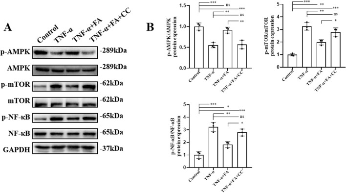
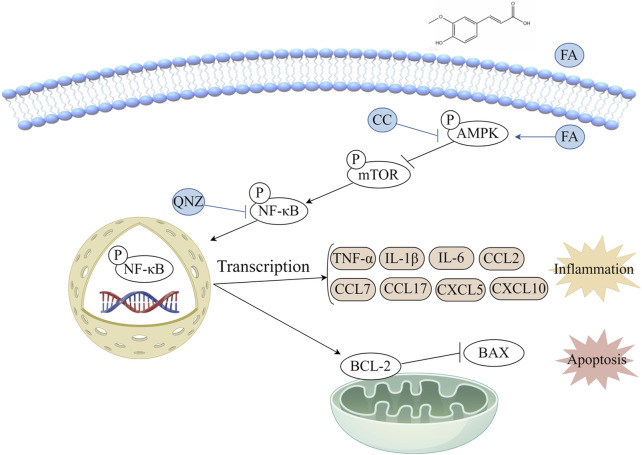
Results: Our in vivo results demonstrated that FA exerted anti-inflammatory effects on KD by inhibiting the infiltration of CD45-positive leukocytes and fibrosis around the coronary artery. Additionally, FA downregulated the levels of inflammatory and chemotactic cytokines, alleviated splenomegaly, and exhibited anti-apoptotic effects on KD by reducing TUNEL-positive cells, downregulating BAX expression, and upregulating BCL-2 expression. In addition, Our in vitro findings showed that FA could effectively inhibit TNF-α-induced HUVEC inflammation like NF-κB inhibitor QNZ by downregulating the expression of pro-inflammatory cytokines as well as attenuated TNF-α-induced HUVEC apoptosis by reducing apoptotic cell numbers and the BAX/BCL-2 ratio, which could be reversed by the AMPK inhibitor compound c (CC). The further mechanistic study demonstrated that FA could restrain vascular endothelial cell inflammation and apoptosis in KD through activating the AMPK/mTOR/NF-κB pathway. However, FA alone is hard to completely restore KD into normal condition.

Conclusion: In conclusion, FA has potential protective effects on KD, suggesting its promising role as an adjuvant for KD therapy in the future.

Keywords: apoptosis; coronary artery; ferulic acid (FA); inflammation; kawasaki disease (KD); vasculitis.


Conflict of interest statement

The authors declare that the research was conducted in the absence of any commercial or financial relationships that could be construed as a potential conflict of interest.

Figures

FIGURE 1
FIGURE 1
FA alleviated coronary artery inflammation in CAWS-induced KD mice. (A) The chemical structure of FA. (B) A flow diagram illustrating the experimental procedure for inducing Kawasaki disease (KD) in mice and treating them with FA. (C) HE staining of around mouse coronary artery in different groups. Scale bars 625 μm, 200 μm. (D) The quantification of HE staining in different groups (n = 3). (E) Immunohistochemistry of CD45 in different groups. Scale bars 625 μm, 200 μm. (F) The quantification of CD45 immunohistochemistry in different groups (n = 3). (G) Mason staining around mouse coronary artery in different groups. Scale bars 400, 200 μm. (H) The quantification of Mason staining in different groups (n = 3). Mean ± SD, * p < 0.05, ** p < 0.01, *** p < 0.001 indicate significant differences, and ns > 0.05 means no significance difference.
FIGURE 2
FIGURE 2
FA decreased inflammation in CAWS-induced KD mice. (A) The representative images of mouse spleens in different groups on day 28. (B) Spleen weight, body weight, and the ratio of spleen weight/body weight on day 28 in different groups (n = 3). (C) The mRNA expression levels of inflammatory cytokines and chemokines in the aortic root and proximal coronary artery regions of mouse heart tissues in each group were evaluated using RT-qPCR (n = 3). (D) Western blot analysis was employed to assess the expression of CD45, IL-1β, and IL-6 proteins in the aortic root and proximal coronary artery regions of mouse heart tissues in each group. (E) The quantification of western blot in different groups (n = 3). Mean ± SD, * p < 0.05, ** p < 0.01, *** p < 0.001 indicate significant differences, and ns > 0.05 means no significance difference.
FIGURE 3
FIGURE 3
FA inhibited vascular endothelial cell apoptosis of coronary arteries in CAWS-KD mice. (A) TUNEL and CD31 staining of paraffin sections in different groups. Scale bars 50 μm. (B) The quantification of TUNEL-positive vascular endothelial cells in different groups (n = 3). (C) The expression levels of BAX and BCL-2 proteins in cardiac tissues of mice in different experimental groups were evaluated using Western blot analysis. (D) The quantification of BAX and BCL-2 expressions in different groups (n = 3). Mean ± SD, * p < 0.05, ** p < 0.01, *** p < 0.001 indicate significant differences, and ns > 0.05 means no significance difference.
FIGURE 4
FIGURE 4
FA had protective effects on CAWS-KD mice through activating AMPK/mTOR/NF-κB pathway. (A) Western blot analysis was employed to assess the expression of relevant biomarkers along the AMPK/mTOR/NF-κB pathway in the aortic root and proximal coronary artery regions of mouse heart tissues in different groups. (B) The statistical analysis of western blot in different groups (n = 3). Mean ± SD, * p < 0.05, ** p < 0.01, *** p < 0.001 indicate significant differences, and ns > 0.05 means no significance difference.
FIGURE 5
FIGURE 5
FA alleviated cell inflammation of TNF-α-induced HUVECs through inhibiting NF-κB pathway. (A) Immunofluorescence staining of IL-1β in each group. Scale bars 50 μm. (B) The quantification of protein levels in each group (n = 3). (C) The gene expression levels of inflammatory cytokines such as IL-1β and IL-6 as well as chemokine CXCL10 in different groups by RT-qPCR (n = 3). (D) The expressions of proteins such as NF-κB, p-NF-κB, IL-1β, and IL-6 in different groups by western blot. (E) The statistical analysis of western blot gel bands in different groups (n = 3). (F) The protein expressions of BAX and BCL-2 in different groups by western blot. (G) The quantification of western blot in different groups (n = 3). Mean ± SD, * p < 0.05, ** p < 0.01, *** p < 0.001 indicate significant differences, and ns > 0.05 means no significance difference.
FIGURE 6
FIGURE 6
FA alleviated cell inflammation of TNF-α-induced HUVECs. (A) Immunofluorescence staining of IL-6 in different groups. Scale bars 50 μm. (B) The statistical analysis of IL-6 fluorescence intensity in different groups (n = 3). (C) Western blot gel images of IL-1β. (D) The bar graphs represent the quantification of protein levels in each group (n = 3). Mean ± SD, * p < 0.05, ** p < 0.01, *** p < 0.001 indicate significant differences, and ns > 0.05 means no significance difference.
FIGURE 7
FIGURE 7
FA alleviated cell apoptosis of TNF-α-induced HUVECs. (A) TUNEL staining of HUVECs in different groups. Scale bars 50 μm. (B) The statistical analysis of TUNEL-positive cells in different groups (n = 3). (C) The apoptosis of HUVECs in different groups by flow cytometry. (D) The statistical analysis of flow cytometry in different groups (n = 3). (E) The protein expressions of BAX and BCL-2 in different groups using western blot. (F) The statistical analysis of BAX and BCL-2 levels in different groups (n = 3). Mean ± SD, * p < 0.05, ** p < 0.01, *** p < 0.001 indicate significant differences, and ns > 0.05 means no significance difference.
FIGURE 8
FIGURE 8
FA exerted protective effects on TNF-α-induced HUVECs through activating AMPK/mTOR/NF-κB pathway. (A) The protein expressions of the associated indicators of AMPK/mTOR/NF-κB pathway in different groups by western blot. (B) The quantification of western blot in different groups (n = 3). Mean ± SD, * p < 0.05, ** p < 0.01, *** p < 0.001 indicate significant differences, and ns > 0.05 means no significance difference.
FIGURE 9
FIGURE 9
Diagram of the potential mechanism involved in the protective effects of FA on KD.

Similar articles

Cited by

References

    1. Ahmad A., Biersack B., Li Y., Kong D., Bao B., Schobert R., et al. (2013). Targeted regulation of PI3K/Akt/mTOR/NF-κB signaling by indole compounds and their derivatives: mechanistic details and biological implications for cancer therapy. Anticancer Agents Med. Chem. 13 (7), 1002–1013. 10.2174/18715206113139990078 - DOI - PMC - PubMed
    1. Babbar R., Dhiman S., Grover R., Kaur A., Arora S. (2021). A comprehensive review on therapeutic applications of ferulic acid and its novel analogues: a brief literature. Mini-Rev Med. Chem. 21 (12), 1578–1593. 10.2174/1389557521666210120111702 - DOI - PubMed
    1. Chen S., Cheng A. C., Wang M. S., Peng X. (2008). Detection of apoptosis induced by new type gosling viral enteritis virus in vitro through fluorescein annexin V-FITC/PI double labeling. World J. Gastroentero. 14 (14), 2174–2178. 10.3748/wjg.14.2174 - DOI - PMC - PubMed
    1. Chen X., Zhou X., Cheng X., Lin L., Wang Q., Zhan R., et al. (2023). Protective effect of ferulic acid on lipopolysaccharide-induced BV2 microglia inflammation via AMPK/mTOR signaling pathway. Molecules 28 (8), 3482. 10.3390/molecules28083482 - DOI - PMC - PubMed
    1. Chen Y., Xue F., Han C., Yang H., Han L., Li K., et al. (2019). Ferulic acid ameliorated placental inflammation and apoptosis in rat with preeclampsia. Clin. Exp. Hypertens. 41 (6), 524–530. 10.1080/10641963.2018.1516773 - DOI - PubMed

LinkOut - more resources