

Expression of vascular endothelial growth factor receptor-2, epidermal growth factor receptor, cyclooxygenase-2, survivin, E-cadherin and Ki-67 in canine nasal carcinomas and sarcomas - a pilot study
- PMID: 39268521
- PMCID: PMC11391428
- DOI: 10.3389/fvets.2024.1388493
Expression of vascular endothelial growth factor receptor-2, epidermal growth factor receptor, cyclooxygenase-2, survivin, E-cadherin and Ki-67 in canine nasal carcinomas and sarcomas - a pilot study
Abstract
Background: Malignant (intra-) nasal tumors (NTs) are the most common cause of chronic nasal discharge in dogs. Besides radiation therapy, palliative therapy is necessary in some dogs. Therefore, studies on receptor expression have supported the utility of tyrosine kinase inhibitors (TKI) in dogs with nasal carcinomas. However, studies on receptor expression in nasal sarcomas are lacking.
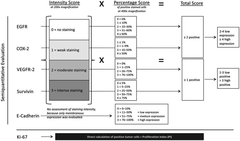
Materials and methods: This study evaluated the expression of vascular endothelial growth factor receptor-2 (VEGFR-2), epidermal growth factor receptor (EGFR), cyclooxigenase-2 (COX-2), Ki-67, survivin and E-cadherin in nasal carcinomas and sarcomas and compared it with tumor (T) categories based on computed tomography (CT).
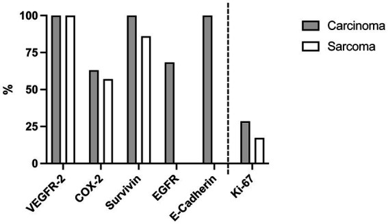
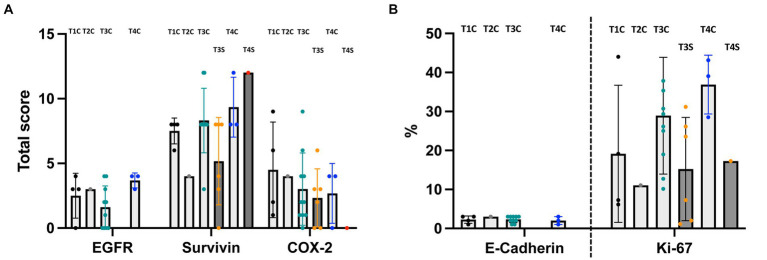
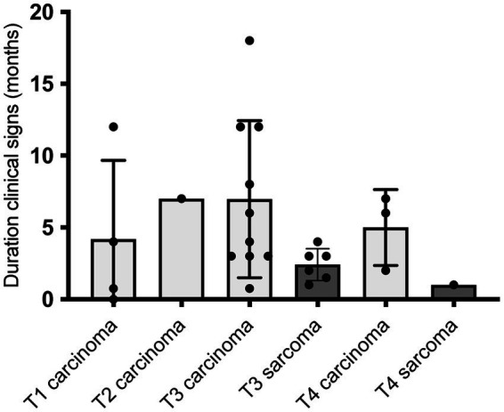
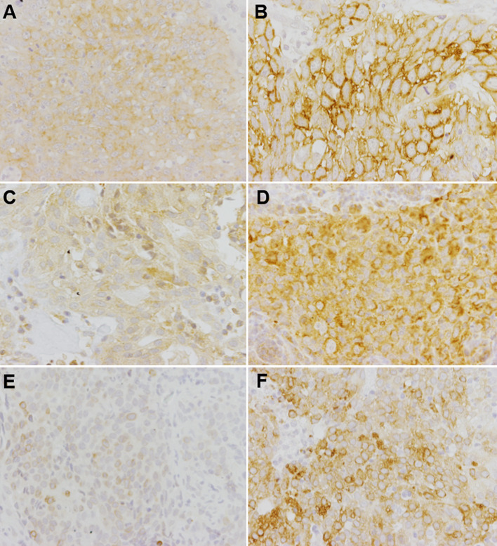
Results: In 26 dogs with NTs, cross sectional imaging and upper airway endoscopy with guided biopsy collection were performed, followed by histopathological examination of NTs, revealing 19 epithelial and 7 mesenchymal tumors. While EGFR and E-cadherin were only expressed by carcinomas, the following markers were expressed by both carcinomas and sarcomas without significant differences between tumor types and T-categories: VEGFR-2 (carcinomas and sarcomas 100%), COX-2 (carcinomas 63%, sarcomas 57%), survivin (carcinomas 100%, sarcomas 86%) and Ki-67 (median expression of 28.5% in carcinomas and 17.3% in sarcomas).
Conclusion: Based on similarities in marker expression between canine carcinomas and sarcomas, clinical studies should further elucidate the use of TKI or COX-2 inhibitors as additional therapy in dogs with nasal sarcomas.
Keywords: T-categories; chronic nasal discharge; computed tomography; cyclooxygenase inhibitors; dogs; immunohistochemistry; nasal tumors; tyrosine kinase inhibitors.
Copyright © 2024 Pauly, Junginger, Oechtering, Hewicker-Trautwein and Rösch.
Conflict of interest statement
The authors declare that the research was conducted in the absence of any commercial or financial relationships that could be construed as a potential conflict of interest.
Figures
Similar articles
-
Diagnostic value of serum survivin, Ki-67 and thymidine kinase in dogs with nasal cavity disease.Front Vet Sci. 2025 Apr 7;12:1553551. doi: 10.3389/fvets.2025.1553551. eCollection 2025. Front Vet Sci. 2025. PMID: 40260215 Free PMC article.
-
Comparison of epidermal growth factor receptor (EGFR) and cyclooxygenase-2 (COX-2) immunohistochemical expression and outcomes in canine nasal carcinomas treated with radiation therapy.J Vet Med Sci. 2022 Sep 5;84(9):1237-1243. doi: 10.1292/jvms.22-0106. Epub 2022 Jul 15. J Vet Med Sci. 2022. PMID: 35851267 Free PMC article.
-
Immunohistochemistry Screening of Different Tyrosine Kinase Receptors in Canine Solid Tumors-Part I: Proposal of a Receptor Panel to Predict Therapies.Int J Mol Sci. 2024 Aug 2;25(15):8438. doi: 10.3390/ijms25158438. Int J Mol Sci. 2024. PMID: 39126006 Free PMC article.
-
Combination antiangiogenic therapy and radiation in head and neck cancers.Oral Oncol. 2014 Jan;50(1):19-26. doi: 10.1016/j.oraloncology.2013.10.003. Epub 2013 Oct 23. Oral Oncol. 2014. PMID: 24269532 Review.
-
Research progress of good markers for canine mammary carcinoma.Mol Biol Rep. 2023 Dec;50(12):10617-10625. doi: 10.1007/s11033-023-08863-x. Epub 2023 Nov 9. Mol Biol Rep. 2023. PMID: 37943402 Review.
Cited by
-
Diagnostic Utility of Canine C-Reactive Protein, Haptoglobin, and 25-Hydroxyvitamin-D in Dogs with Nasal Cavity Disease.Animals (Basel). 2024 Oct 9;14(19):2908. doi: 10.3390/ani14192908. Animals (Basel). 2024. PMID: 39409857 Free PMC article.
-
Diagnostic value of serum survivin, Ki-67 and thymidine kinase in dogs with nasal cavity disease.Front Vet Sci. 2025 Apr 7;12:1553551. doi: 10.3389/fvets.2025.1553551. eCollection 2025. Front Vet Sci. 2025. PMID: 40260215 Free PMC article.
-
Computed Tomography-Guided Radiofrequency Ablation of Nasal Carcinomas in Dogs.Animals (Basel). 2024 Dec 20;14(24):3682. doi: 10.3390/ani14243682. Animals (Basel). 2024. PMID: 39765586 Free PMC article.
References
-
- Madewell B, Priester W, Gillette E, Snyder S. Neoplasms of the nasal passages and paranasal sinuses in domesticated animals as reported by 13 veterinary colleges. Am J Vet Res. (1976) 37:851–6. PMID: - PubMed
-
- Kleiter M. Tumoren des Respirationstrakts-Tumoren der Nasenhöhle und Nasennebenhöhlen In: Kessler M, editor. Kleintieronkologie-Diagnose und Therapie von Tumorerkrankungen bei Hund und Katze. 3rd ed. Stuttgart, Germany: Enke; (2013). 305–23.
-
- Patnaik A. Canine sinonasal neoplasms: clinicopathological study of 285 cases. J Am Anim Hosp Assoc. (1989) 25:103–14.
LinkOut - more resources
Full Text Sources
Research Materials
Miscellaneous