. 2024 Oct 1;17(10):dmm050852.
doi: 10.1242/dmm.050852. Epub 2024 Oct 9.

Functional cardiac consequences of β-adrenergic stress-induced injury in a model of Duchenne muscular dystrophy

Affiliations

Functional cardiac consequences of β-adrenergic stress-induced injury in a model of Duchenne muscular dystrophy

Conner C Earl et al. Dis Model Mech. .

Abstract

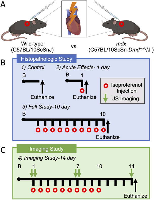
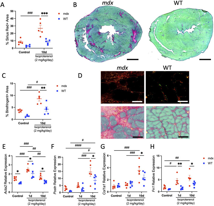
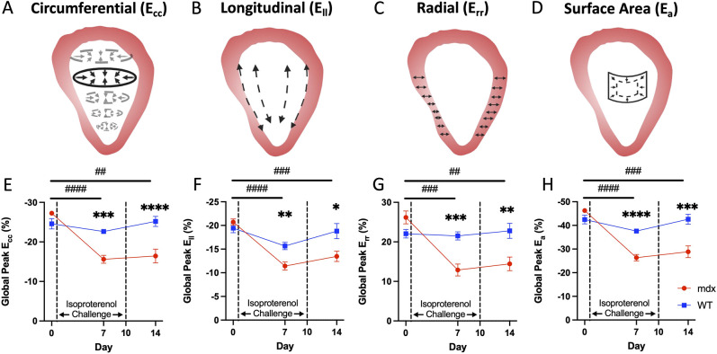
Cardiomyopathy is the leading cause of death in Duchenne muscular dystrophy (DMD); however, in the mdx mouse model of DMD, the cardiac phenotype differs from that seen in DMD-associated cardiomyopathy. Although some have used pharmacologic stress to stimulate injury and enhance cardiac pathology in the mdx model, many methods lead to high mortality with variable cardiac outcomes, and do not recapitulate the structural and functional cardiac changes seen in human disease. Here, we describe a simple and effective method to enhance the cardiac phenotype model in mdx mice using advanced 2D and 4D high-frequency ultrasound to monitor cardiac dysfunction progression in vivo. mdx and wild-type mice received daily low-dose (2 mg/kg/day) isoproterenol injections for 10 days. Histopathological assessment showed that isoproterenol treatment increased myocyte injury, elevated serum cardiac troponin I levels and enhanced fibrosis in mdx mice. Ultrasound revealed reduced ventricular function, decreased wall thickness, increased volumes and diminished cardiac reserve in mdx compared to wild-type mice. Our findings highlight the utility of challenging mdx mice with low-dose isoproterenol as a valuable model for exploring therapies targeting DMD-associated cardiac pathologies.

Keywords: mdx; 4DUS; Cardiac strain; Duchenne muscular dystrophy; Isoproterenol; Mouse model.


Conflict of interest statement

Competing interests C.J.G. is a paid consultant of FUJIFILM VisualSonics Inc.

Figures

Fig. 1.
Fig. 1.
Study overview. (A) Schematic representing subcutaneous injection of 2 mg/kg/day isoproterenol to induce β-adrenergic cardiac stimulation and injury. (B) Histopathological study timeline. (C) Imaging study timeline. US, ultrasound.
Fig. 2.
Fig. 2.
Isoproterenol promotes sarcolemmal injury in dystrophin-deficient cardiac myocytes. The acute and chronic effects of isoproterenol (2 mg/kg/day) were assessed in mdx and wild-type (WT) mice treated for 1 (n=10 per group) or 10 (n=5 per group) days. Control mice were injected with an equal volume of saline for 10 days (n=5 mdx and n=4 WT). (A,B) To assess sarcolemmal damage, mid-chamber coronal sections of ventricles were immunolabeled with anti-IgM (red) and counterstained with wheat germ agglutinin (WGA; green) to visualize tissue morphology. (A) Cardiac injury was detected transiently in mdx mice after isoproterenol challenge, with IgM+ cardiac myocytes occupying ∼8.5% of the myocardial area after a single isoproterenol treatment. Cardiac myocyte injury was scarcely detected in mdx mice after chronic isoproterenol administration or in WT mice across all conditions. (B) Representative mdx (top row) and WT (bottom row) whole-ventricle cross-section and high-magnification images show prominent areas of injury (red) after acute isoproterenol treatment in mdx mice. Scale bars: 500 µm. Insets: high-magnification images confirm that the IgM+ signal is localized to cardiac myocytes in mdx mice. Scale bars: 50 µm. (C) Serum cardiac troponin I (cTnI) levels were measured by ELISA as an independent assay of cardiac injury. Serum cTnI levels were also transiently elevated in mdx mice after acute isoproterenol treatment (n=5 per group). Data are presented as mean±s.e.m. All P-values are based on two-way ANOVA with Tukey's multiple comparison test. **P<0.01 and ***P<0.001 versus WT within a treatment condition. ##P<0.01 and ###P<0.001 between treatment groups within a genotype. d, day.
Fig. 3.
Fig. 3.
Distinct cardiac growth and stress responses to β-adrenergic stimulation in dystrophic hearts. Heart weight normalized to tibia length (HW/TL) and heart weight normalized to body mass (HW/BM) were used as indices of cardiac growth in mdx and WT mice treated for 1 (n=10 per group) or 10 (n=5 per group) days with isoproterenol, and control mice (n=5 mdx and n=4 WT). (A) HW/TL ratio was higher in mdx mice than in WT mice in all conditions. Chronic isoproterenol treatment resulted in an increased HW/TL ratio in WT mice only. (B) HW/BM ratio increased with chronic isoproterenol treatment in WT mice relative to that in control and mdx mice. (C) Control mdx mice have a higher mean minimum ferret diameter than that of WT mice. WT mice treated with isoproterenol for 10 days show significant hypertrophy of cardiac myocytes. (D) Representative images of mid-chamber transverse cardiac sections stained with WGA to visualize cell borders. Scale bars: 20 µm. (E,F) Quantitative PCR (QPCR) analyses of mdx and WT ventricles expressed as mRNA levels relative to those of WT control. Nppa (E) and Nppb (F) mRNA expression increased in mdx mice with chronic isoproterenol challenge and relative to that of WT. (G) Ventricular wall thickness was measured at key time points by ultrasound (n=10 per genotype). At baseline, wall thickness was greater in mdx hearts than in WT hearts. However, in response to isoproterenol treatment, wall thickness decreased in mdx mice, becoming less thick than in WT mice at day 14. (H) Mid-chamber coronal sections of mdx (left) and WT (right) ventricles stained with Masson's trichrome show characteristic fibrosis and thinning of the free wall in mdx mice at day 14. Scale bars: 1 mm. Data are presented as mean±s.e.m. All P-values are based on two-way ANOVA with Tukey's multiple comparison test. *P<0.05, **P<0.01, ***P<0.001 and ****P<0.0001 versus WT within a treatment condition. #P<0.05, ##P<0.01, ###P<0.001 and ####P<0.0001 between treatment groups within a genotype.
Fig. 4.
Fig. 4.
Isoproterenol promotes fibrotic replacement of the dystrophic myocardium. (A-G) Fibrotic area was measured on mid-chamber cross-sections stained with Sirius Red Fast Green (SRFG) in control (n=5 mdx and n=4 WT) and chronic isoproterenol-treated (n=5 per genotype) mice. (A,B) The area of the myocardium occupied by collagen (red stain) is increased in mdx mice (left in B) after chronic isoproterenol challenge relative to that in control and isoproterenol-treated WT mice (right in B). Scale bars: 1 mm. (C,D) Collagen density was visualized by polarized light microscopy in isoproterenol-treated mdx and WT mice. (C) Chronic isoproterenol treatment increased areas of birefringence in mdx and WT mice. (D) Fibrotic lesions of chronic isoproterenol-treated mdx mice contained prominent birefringent areas (red and orange), indicating the presence of densely bundled collagen fibers (top row). Representative overlay images of brightfield and polarized light mdx and WT ventricles with birefringent areas pseudo-colored yellow to enhance image contrast in image overlays (bottom row). Scale bars: 50 μm. (E-H) QPCR analyses of transcripts encoding markers of activated fibroblasts (E,F) and connective tissue proteins (G,H). (E) Acta2 mRNA levels were elevated in mdx mice relative to those in WT mice at baseline and with isoproterenol stimulation. (F) Induction of Postn transcripts was greater with isoproterenol stimulation in mdx mice than in WT mice. (G,H) Genes encoding extracellular matrix components Col1a1 (G) and Fn1 (H) were further increased in mdx mice relative to those in WT mice with isoproterenol treatment. Data are expressed as mRNA levels relative to those of the WT control group and presented as mean±s.e.m. All P-values are based on two-way ANOVA with Tukey's multiple comparison test. *P<0.05, **P<0.01 and ***P<0.001 versus WT within a treatment condition. #P<0.05, ##P<0.01, ###P<0.001 and ####P<0.0001 between treatment groups within a genotype.
Fig. 5.
Fig. 5.
Cardiac function decreases with isoproterenol injury in dystrophic hearts. (A) Left-ventricular ejection fraction (LVEF) in mdx mice was significantly reduced by day 7 and further reduced at day 14. (B,C) The left-ventricular end-diastolic volume (EDV; B) and end-systolic volume (ESV; C) in mdx mice were significantly greater than those in WT mice at day 14, signifying left-ventricular dilation. (D,E) There were no significant changes in the cardiac output (CO; D) or heart rate (HR; E) in either the WT or mdx group. (F) 2D ultrasound parasternal long-axis images at peak systole for both mdx and WT mice at day 0, day 7 and day 14 with LVEF label (EF). Scale bar: 1 mm. These findings suggest that the mdx mice exhibited dilated cardiomyopathy by day 14 when given an isoproterenol challenge. Data are presented as mean±s.e.m. All P-values are based on two-way ANOVA with Tukey's multiple comparison test. *P<0.05, **P<0.01 and ***P<0.001 versus WT at a specified timepoint. #P<0.05, ##P<0.01 and ###P<0.001 mdx difference between timepoints. bpm, beats/min.
Fig. 6.
Fig. 6.
Cardiac response to isoproterenol challenge decreases over time in dystrophic hearts. (A,B) The initial compensatory response to an isoproterenol administration measured at exactly 1 min before and 1 min after the injection was apparent in the increase in HR and LVEF in both mdx and WT mice. (C) In the 2D ultrasound images of the left ventricle, this compensation can be seen as endocardial walls coming close together during systole. (D,E) After 7 days of exposure to isoproterenol injury, this compensatory response is impaired in the mdx mice, in which the ΔLVEF (D) and ΔHR (E) are reduced in comparison to those in WT. (F) There is little to no qualitative change in the left-ventricular chamber in the mdx mice compared to WT mice immediately after isoproterenol injection. After prolonged exposure to an isoproterenol challenge, the ability of mdx mice to compensate for a single injection is impaired in comparison to the robust compensation of WT mice. Data are presented as mean±s.e.m. All P-values are based on two-way ANOVA with Tukey's multiple comparison test. **P<0.01 and ****P<0.0001 versus WT. ##P<0.01, ###P<0.001 and ####P<0.0001 difference between pre- and post-injection.
Fig. 7.
Fig. 7.
4D strain magnitude decreases in dystrophic hearts with isoproterenol injury. (A-D) Global circumferential, longitudinal, radial and surface area strain were calculated with 4D ultrasound (4DUS) measurements. (E-H) In the mdx mice with isoproterenol injury, the magnitude of strain was significantly lowered by day 7. There was a slight recovery in ventricular strain by day 14, yet the strain magnitude remains significantly reduced. 4D strain decreases in dystrophic hearts when given an isoproterenol challenge. After the completion of the isoproterenol challenge, there is some functional recovery, but irreversible damage remains. Data are presented as mean±s.e.m. All P-values are based on two-way ANOVA with Tukey's multiple comparison test. *P<0.05, **P<0.01, ***P<0.001 and ****P<0.0001 versus WT at a specified timepoint. ##P<0.01, ###P<0.001 and ####P<0.0001 mdx difference between timepoints. Ecc, circumferential strain; Ell, longitudinal strain; Err, radial strain; Ea, surface area strain.

Update of

Similar articles

Cited by

References

    1. Au, C. G., Butler, T. L., Sherwood, M. C., Egan, J. R., North, K. N. and Winlaw, D. S. (2011). Increased connective tissue growth factor associated with cardiac fibrosis in the mdx mouse model of dystrophic cardiomyopathy. Int. J. Exp. Pathol. 92, 57-65. 10.1111/j.1365-2613.2010.00750.x - DOI - PMC - PubMed
    1. Betts, C. A., Saleh, A. F., Carr, C. A., Hammond, S. M., Coenen-Stass, A. M. L., Godfrey, C., Mcclorey, G., Varela, M. A., Roberts, T. C., Clarke, K.et al. (2015). Prevention of exercised induced cardiomyopathy following Pip-PMO treatment in dystrophic mdx mice. Sci. Rep. 5, 8986. 10.1038/srep08986 - DOI - PMC - PubMed
    1. Birnkrant, D. J., Bushby, K., Bann, C. M., Alman, B. A., Apkon, S. D., Blackwell, A., Case, L. E., Cripe, L., Hadjiyannakis, S., Olson, A. K.et al. (2018a). Diagnosis and management of Duchenne muscular dystrophy, part 2: respiratory, cardiac, bone health, and orthopaedic management. Lancet Neurol. 17, 347-361. 10.1016/S1474-4422(18)30025-5 - DOI - PMC - PubMed
    1. Birnkrant, D. J., Bushby, K., Bann, C. M., Apkon, S. D., Blackwell, A., Colvin, M. K., Cripe, L., Herron, A. R., Kennedy, A., Kinnett, K.et al. (2018b). Diagnosis and management of Duchenne muscular dystrophy, part 3: primary care, emergency management, psychosocial care, and transitions of care across the lifespan. Lancet Neurol. 17, 445-455. 10.1016/S1474-4422(18)30026-7 - DOI - PMC - PubMed
    1. Boluyt, M. O., Long, X., Eschenhagen, T., Mende, U., Schmitz, W., Crow, M. T. and Lakatta, E. G. (1995). Isoproterenol infusion induces alterations in expression of hypertrophy-associated genes in rat heart. Am. J. Physiol. 269, H638-H647. 10.1152/ajpheart.1995.269.2.H638 - DOI - PubMed

MeSH terms