No replicating evidence for anti-amyloid-β autoantibodies in cerebral amyloid angiopathy-related inflammation
- PMID: 39268830
- PMCID: PMC11514902
- DOI: 10.1002/acn3.52169
No replicating evidence for anti-amyloid-β autoantibodies in cerebral amyloid angiopathy-related inflammation
Abstract
Objective: Elevated levels of anti-amyloid-β (anti-Aβ) autoantibodies in cerebrospinal fluid (CSF) have been proposed as a diagnostic biomarker for cerebral amyloid angiopathy-related inflammation (CAA-RI). We aimed to independently validate the immunoassay for quantifying these antibodies and evaluate its diagnostic value for CAA-RI.
Methods: We replicated the immunoassay to detect CSF anti-Aβ autoantibodies using CSF from CAA-RI patients and non-CAA controls with unrelated disorders and further characterized its performance. Moreover, we conducted a literature review of CAA-RI case reports to investigate neuropathological and CSF evidence of the nature of the inflammatory reaction in CAA-RI.
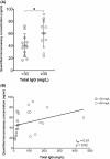
Results: The assay demonstrated a high background signal in CSF, which increased and corresponded with higher total immunoglobulin G (IgG) concentration in CSF (rsp = 0.51, p = 0.02). Assay levels were not elevated in CAA-RI patients (n = 6) compared to non-CAA controls (n = 20; p = 0.64). Literature review indicated only occasional presence of B-lymphocytes and plasma cells (i.e., antibody-producing cells), alongside the abundant presence of activated microglial cells, T-cells, and other monocyte lineage cells. CSF analysis did not convincingly indicate intrathecal IgG production.
Interpretation: We were unable to reproduce the reported elevation of anti-Aβ autoantibody concentration in CSF of CAA-RI patients. Our findings instead support nonspecific detection of IgG levels in CSF by the assay. Reviewed CAA-RI case reports suggested a widespread cerebral inflammatory reaction. In conclusion, our findings do not support anti-Aβ autoantibodies as a diagnostic biomarker for CAA-RI.
© 2024 The Author(s). Annals of Clinical and Translational Neurology published by Wiley Periodicals LLC on behalf of American Neurological Association.
Conflict of interest statement
The authors declare that they have no conflicts of interest to report.
Figures


References
-
- Piazza F, Greenberg SM, Savoiardo M, et al. Anti‐amyloid β autoantibodies in cerebral amyloid angiopathy‐related inflammation: implications for amyloid‐modifying therapies. Ann Neurol. 2013;73(4):449‐458. - PubMed
-
- Chung KK, Anderson NE, Hutchinson D, Synek B, Barber PA. Cerebral amyloid angiopathy related inflammation: three case reports and a review. J Neurol Neurosurg Psychiatry. 2011;82(1):20‐26. - PubMed
-
- Auriel E, Charidimou A, Gurol ME, et al. Validation of clinicoradiological criteria for the diagnosis of cerebral amyloid angiopathy‐related inflammation. JAMA Neurol. 2016;73(2):197‐202. - PubMed
MeSH terms
Substances
Grants and funding
- R01 NS104147/NS/NINDS NIH HHS/United States
- Dutch Brain Foundation
- 2019 T060/Dutch Heart Foundation
- Stryker
- LSHM17016/Top Sector Life Sciences & Health
- NR170024/The Galen and Hilary Weston Foundation
- WE.03-2022-17/Alzheimer Nederland
- Cerenovus
- HA2015.01.06/Brain Foundation Netherlands
- Medtronic
- P2021-18/ParkinsonNL
- National Health Care Institute
- 5R01NS104147-02/National Institutes of Health, USA
- ART-EXT2010-1/Alzheimer's Research UK
- ART/PG2006/4/Alzheimer's Research UK
- G501033/Medical Research Council UK
- Stichting Alkemade Keuls-Fonds
- 2021038368/ZONMW_/ZonMw/Netherlands
- 10510032120006/ZONMW_/ZonMw/Netherlands
- 10510032120003/ZONMW_/ZonMw/Netherlands
- 733050822/ZONMW_/ZonMw/Netherlands
- 2021-079/Maag-Lever-Darm Stichting
- CVON2015-01: CONTRAST/Dutch Heart Foundation
LinkOut - more resources
Full Text Sources

