. 2024 Sep;4(9):e70013.
doi: 10.1002/cpz1.70013.

Set Up and Utilization of a Three-Dimensional In Vitro Bioreactor System for Human Intestinal Studies and Microbial Co-Cultures

Affiliations

Set Up and Utilization of a Three-Dimensional In Vitro Bioreactor System for Human Intestinal Studies and Microbial Co-Cultures

Sara E Rudolph et al. Curr Protoc. 2024 Sep.

Abstract

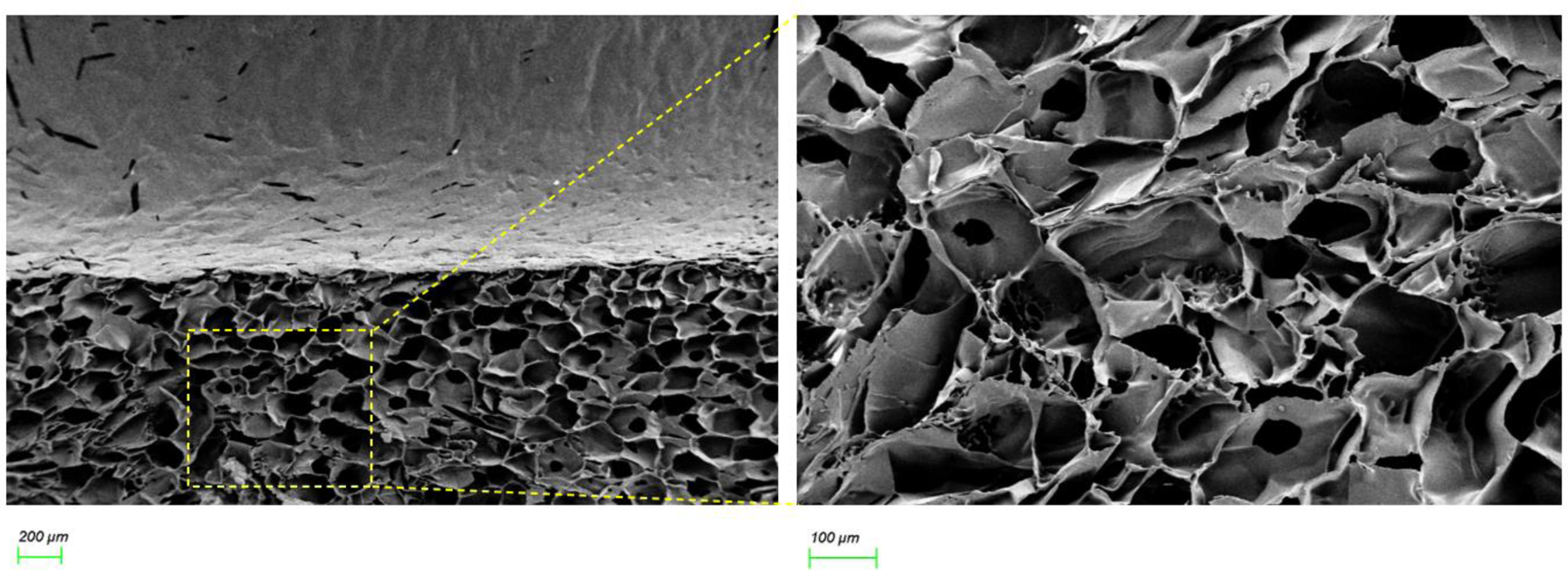
The study of human intestinal physiology and host-microbe interactions is crucial for understanding gastrointestinal health and disease. Traditional two-dimensional cell culture models lack the complexity of the native intestinal environment, limiting their utility in studying intestinal biology. Here, we present a detailed protocol for the set up and utilization of a three-dimensional (3D) in vitro bioreactor system for human intestinal studies and bacterial co-culture. This article outlines the design and assembly of the bioreactor system, scaffold fabrication, bacterial culture techniques, analysis methods, and troubleshooting tips. By providing step-by-step instructions, the goal is to enable other laboratories to utilize physiologically relevant tissue models of the human intestine, incorporating key features, such as nutrient flow, multiple human cell types, 3D architecture, and microbial communities. The incorporation of commensal bacteria into the bioreactor system allows for the investigation of complex host-microbe interactions, providing insight into gastrointestinal health and pathology. This article serves as a comprehensive resource for scientists seeking to advance their understanding of intestinal biology toward the development of novel therapeutic strategies for gastrointestinal disorders. © 2024 Wiley Periodicals LLC. Basic Protocol 1: Scaffold design Basic Protocol 2: Intestinal cell culture: Caco2 cells Basic Protocol 3: Intestinal cell culture: organoids Basic Protocol 4: Bioreactor design and set up Basic Protocol 5: Bacteria in 3D bioreactor set up Basic Protocol 6: Bacteria and drug dosing.

Keywords: 3D silk scaffolds; bacteria; bioreactor; intestinal epithelium; organoids.


Conflict of interest statement

Conflict of Interest: The authors declare no conflict of interest.

Figures

Figure 1:
Figure 1:
Overview of the Protocols - This diagram provides a visual summary of the steps and methodologies detailed in the protocol paper. Each image represents a basic protocol which will be described in detail in this paper.
Figure 2:
Figure 2:
Scanning electron microscopy (SEM) images of final silk sponge scaffolds demonstrating a smooth luminal space and a bulk space with interconnected pores.
Figure 3:
Figure 3:
Full silk processing and scaffold fabrication process. All sets in the silk processing and scaffold design and construction are shown.
Figure 4:
Figure 4:
Scaffold Seeding and Bioreactor Overview. Digestion and seeding of intestinal epithelial cells is shown along with incorporation of these cells into the scaffold system described in Basic Protocol 3. The full scaffold design is also shown, along with a breakdown of how to introduce bacteria into the system and a final view of the luminal and bulk contents of the scaffold.
Figure 5:
Figure 5:
Schematic of 3D bioreactor setup with both intestinal epithelial cells and a bacterial community.
Figure 6:
Figure 6:
Varying ratios of E. coli Nissle (EcN) and human microbiome. Immunofluorescent staining for tight junctions (zonula occludens 1(ZO-1)), green fluorescent protein (GFP)-positive EcN, and 4′,6-diamidino-2-phenylindole (DAPI) are shown for 4, 24, and 48 hours. SEM images are shown for 24 hr. The human microbiome component is co-cultured up to 24 hours, retaining ZO-1 and colonizing the microvilli of the intestinal epithelium as seen in the SEM images (bottom row). Bacteria appear to colonize more in areas of less dense microvilli.
Figure 7:
Figure 7:
Guanidinoacetic acid (GAA) can be measured by LC-MS in the bulk media compartment of the bioreactor setup following a dose into the luminal compartment. Starting media contains either no GAA, human microbiota, 10μM GAA, or 10μM GAA along with human microbiota. GAA can clearly be detected in the bulk space after 24hrs in both samples containing a luminal dose of GAA.
Figure 8:
Figure 8:
SEM images taken at various time points in both flow and static bioreactor setups. Compared to static, the flow system improves microbiome co-culture as can be seen by the breakdown in tight junctions starting at the 48 hr timepoint for both static cultures with microbiota included.
Figure 9:
Figure 9:
Timetable for full set-up of bioreactor and culturing of cells.

References

    1. Agrawal CM, & Ray RB (2001). Biodegradable polymeric scaffolds for musculoskeletal tissue engineering. J Biomed Mater Res, 55(2), 141–150. doi: 10.1002/1097-4636(200105)55:2<141::aid-jbm1000>3.0.co;2-j - DOI - PubMed
    1. Altay G, Tosi S, Garcia-Diaz M, & Martinez E (2020). Imaging the Cell Morphological Response to 3D Topography and Curvature in Engineered Intestinal Tissues. Front Bioeng Biotechnol, 8, 294. doi: 10.3389/fbioe.2020.00294 - DOI - PMC - PubMed
    1. Altman GH, Diaz F, Jakuba C, Calabro T, Horan RL, Chen J, . . . Kaplan DL (2003). Silk-based biomaterials. Biomaterials, 24(3), 401–416. doi: 10.1016/s0142-9612(02)00353-8 - DOI - PubMed
    1. Aziza A, Mahmoud R, Zahran E, & Gadalla H (2020). Dietary supplementation of guanidinoacetic acid improves growth, biochemical parameters, antioxidant capacity and cytokine responses in Nile tilapia (Oreochromis niloticus). Fish Shellfish Immunol, 97, 367–374. doi: 10.1016/j.fsi.2019.12.052 - DOI - PubMed
    1. Castano AG, Garcia-Diaz M, Torras N, Altay G, Comelles J, & Martinez E (2019). Dynamic photopolymerization produces complex microstructures on hydrogels in a moldless approach to generate a 3D intestinal tissue model. Biofabrication, 11(2), 025007. doi: 10.1088/1758-5090/ab0478 - DOI - PubMed

MeSH terms

LinkOut - more resources