Case Reports
. 2024 Nov;102(11):1343-1353.
doi: 10.1007/s00109-024-02482-0. Epub 2024 Sep 13.

Revising pathogenesis of AP1S1-related MEDNIK syndrome: a missense variant in the AP1S1 gene as a causal genetic lesion

Affiliations
Case Reports

Revising pathogenesis of AP1S1-related MEDNIK syndrome: a missense variant in the AP1S1 gene as a causal genetic lesion

Marketa Rackova et al. J Mol Med (Berl). 2024 Nov.

Abstract

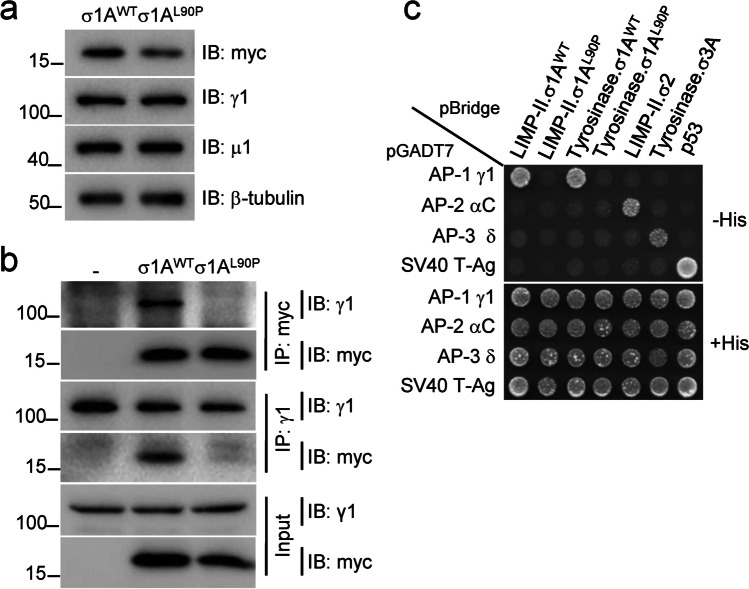
MEDNIK syndrome is a rare autosomal recessive disease characterized by mental retardation, enteropathy, deafness, peripheral neuropathy, ichthyosis, and keratoderma, and caused by variants in the adaptor-related protein complex 1 subunit sigma 1 (AP1S1) gene. This gene encodes the σ1A protein, which is a subunit of the adaptor protein complex 1 (AP-1), a key component of the intracellular protein trafficking machinery. Previous work identified three AP1S1 nonsense, frameshift and splice-site variants in MEDNIK patients predicted to encode truncated σ1A proteins, with consequent AP-1 dysfunction. However, two AP1S1 missense variants (c.269 T > C and c.346G > A) were recently reported in patients who presented with severe enteropathy but no additional symptoms of MEDNIK. This condition was described as a novel non-syndromic form of congenital diarrhea caused specifically by the AP1S1 missense variants. In this study, we report two patients with the same c.269 T > C variant, who, contrary to the previous cases, presented as complete MEDNIK syndrome. These data substantially revise the presentation of disorders associated with AP1S1 gene variants and indicate that all the identified pathogenic AP1S1 variants result in MEDNIK syndrome. We also provide a series of functional analyses that elucidate the impact of the c.269 T > C variant on σ1A function, contributing to a better understanding of the molecular pathogenesis of MEDNIK syndrome. KEY MESSAGES: A missense AP1S1 c.269 T > C (σ1A L90P) variant causes full MEDNIK syndrome. The σ1A L90P variant is largely unable to assemble into the AP-1 complex. The σ1A L90P variant fails to bind [DE]XXXL[LI] sorting motifs. The σ1A L90P variant results in loss-of-function of the protein.

Keywords: AP1S1; Coatopathies; Congenital diarrhea; MEDNIK; Missense variants.


Conflict of interest statement

The authors have no relevant financial or non-financial interests to disclose.

Figures

Fig.1
Fig.1
Structure of the AP-1 complex and genetic analysis. a Schematic representation of the AP-1 complex depicting its γ, β1, μ1 and σ1 subunits. The γ, μ1 and σ1 subunits occur as multiple isoforms encoded by different genes, namely, γ1 and γ2, μ1A and μ1B, and σ1A, σ1B and σ1C, respectively. Also shown are the trunk, hinge and ear domains of γ and β1. The trunk domains of γ and β1 together with the μ1 and σ1 subunits constitute the core of the AP-1 complex. The indicated tyrosine-based YXXØ and dileucine-based [DE]XXXL[LI] sorting signals in the cytosolic tails of transmembrane protein cargos are recognized by the AP-1 μ1 subunit and the γ-σ1 hemicomplex, respectively. b Detection of AP1S1 variant c.269 T > C. Representative sequence traces from patient 1 and her mother and father, and from patient 2 and his mother. c Pedigree of patient 1. Arrow indicates proband, square—male, circle—female, diagonal line—deceased, small triangle—termination of pregnancy
Fig.2
Fig.2
The AP-1 σ1A L90P substitution impairs assembly of the AP-1 complex and recognition of dileucine signals. a Decreased expression of myc-tagged σ1A L90P relative to WT myc-tagged σ1A expressed by transient transfection in HeLa cells and analyzed by SDS-PAGE and immunoblotting (IB). Blots of endogenous γ1, μ1 and β-tubulin are included as loading controls. b Impaired assembly of myc-tagged σ1A L90P into the AP-1 complex. HEK293T cells were transiently transfected with plasmids encoding either WT or L90P myc-tagged σ1A, and cell extracts were subjected to immunoprecipitation (IP) with anti-myc followed by SDS-PAGE and IB with anti-γ1 or anti-myc, or IP with anti-γ1 followed by SDS-PAGE and IB with anti-γ1 or anti-myc. Untransfected cells (-) were used as control. Notice that both permutations of IP and IB showed decreased co-immunoprecipitation of endogenous γ1 with myc-tagged σ1A L90P relative to myc-tagged WT σ1A (ranging from 0 to 13%, depending on the antibody combination; first and fourth blots from top). c Y3H assays showing lack of interaction of σ1A L90P with dileucine-based sorting signals. The AP-1 γ1, AP-2 αC and AP-3 δ subunits were subcloned in the Gal4 transcriptional activation domain (AD) vector pGADT7. The cytosolic tails of LIMP-II or tyrosinase and the indicated σ subunits were subcloned in the MCS1 and MCS2 of the Gal4 DNA binding domain (BD) vector pBridge, respectively. Transformants were plated on medium lacking leucine, tryptophan and methionine but containing histidine (+ His, bottom panel) to control for viability and loading, and on the same medium lacking histidine (-His, top panels) to detect protein interactions. The top panel shows the interaction of the γ1-σ1A hemicomplex with the dileucine motifs in the cytosolic tails of LIMP-II and tyrosinase (ERAPLI and ERQPLL, respectively [6]; and that the σ1A L90P substitution abrogates this interaction. Note the selective interaction of the LIMP-II and tyrosinase tails with the AP-1 γ1-σ1A hemicomplex but not with mismatched combinations of AP subunits. Additional controls in the assay include the interaction of the LIMP-II tail with AP-2 αC-σ2 and of the tyrosinase tail with AP-3 δ-σ3A (but not with mismatched combinations of AP subunits). Yeast co-transformation of pBridge-based constructs with a Gal4 AD-SV40 T-Ag fusion construct and of AD-AP subunit fusions with a Gal4 BD-p53 construct were used as negative controls. Co-transformants co-expressing AD-SV40 T-Ag and BD-p53 fusions provided a positive control for interactions
Fig.3
Fig.3
The σ1A L90P substitution prevents rescue of the association of AP-1 with TGN/endosomes in triple σ1-KO cells. a IB analysis of cell lysates from WT HAP1 cells and triple σ1-KO HAP1 cells untransfected (-) or transfected with plasmids encoding myc-tagged WT or L90P σ1A. Notice the partial rescue of γ1 levels in triple σ1-KO cells expressing σ1WT but not L90P σ1A. IB with anti-β-tubulin is shown as loading control. b Confocal immunofluorescence microscopy of WT and triple σ1-KO HAP1 cells stained for endogenous γ1 (red channel) and the TGN marker TGN46 (green channel). Notice the co-localization of γ1 and TGN46 in the perinuclear region of WT HAP1 cells, and the marked reduction in γ1 signal in the triple σ1-KO cells. c Confocal immunofluorescence microscopy of triple σ1-KO HAP1 cells transfected with plasmids encoding myc-tagged WT or L90P σ1A, and stained for endogenous γ1 (red channel) and the myc epitope (green channel). Notice the rescue of perinuclear γ1 immunostaining by expression of WT but not L90P σ1A. In b and c, cell edges are indicated with dashed lines; scale bars: 10 μm
Fig.4
Fig.4
Immunohistochemical analysis of patient samples. a Immunohistochemical staining of ZO-1 in intestinal biopsy of healthy control and patient 2. Arrows show apical localization of the protein. b Immunohistochemical staining of claudin-3 in intestinal biopsy of healthy control and patient 2. Arrows show basolateral localization of the protein

References

    1. García-Cazorla A, Oyarzábal A, Saudubray JM et al (2022) Genetic disorders of cellular trafficking. Trends Genet 38:724–751. 10.1016/J.TIG.2022.02.012 - PubMed
    1. Dell’Angelica EC, Bonifacino JS (2019) Coatopathies: Genetic Disorders of Protein Coats. Annu Rev Cell Dev Biol 35:131–168. 10.1146/ANNUREV-CELLBIO-100818-125234 - PMC - PubMed
    1. Sanger A, Hirst J, Davies AK, Robinson MS (2019) Adaptor protein complexes and disease at a glance. J Cell Sci 132(20):jcs222992. 10.1242/JCS.222992 - PubMed
    1. Janvier K, Kato Y, Boehm M et al (2003) Recognition of dileucine-based sorting signals from HIV-1 Nef and LIMP-II by the AP-1 gamma-sigma1 and AP-3 delta-sigma3 hemicomplexes. J Cell Biol 163:1281–1290. 10.1083/JCB.200307157 - PMC - PubMed
    1. Doray B, Lee I, Knisely J et al (2007) The gamma/sigma1 and alpha/sigma2 hemicomplexes of clathrin adaptors AP-1 and AP-2 harbor the dileucine recognition site. Mol Biol Cell 18:1887–1896. 10.1091/MBC.E07-01-0012 - PMC - PubMed

Publication types

Substances

LinkOut - more resources