. 2024 Sep 13;13(1):17.
doi: 10.1186/s13619-024-00200-2.

Endogenous retroviral ERVH48-1 promotes human urine cell reprogramming

Affiliations

Endogenous retroviral ERVH48-1 promotes human urine cell reprogramming

Yuling Peng et al. Cell Regen. .

Abstract

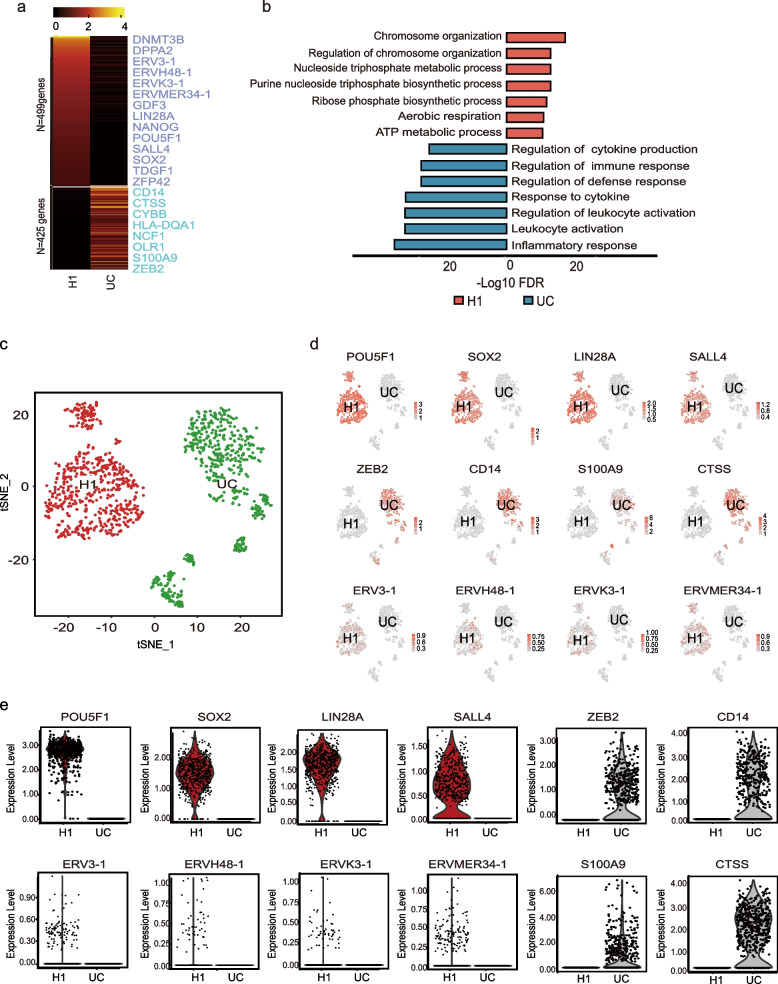
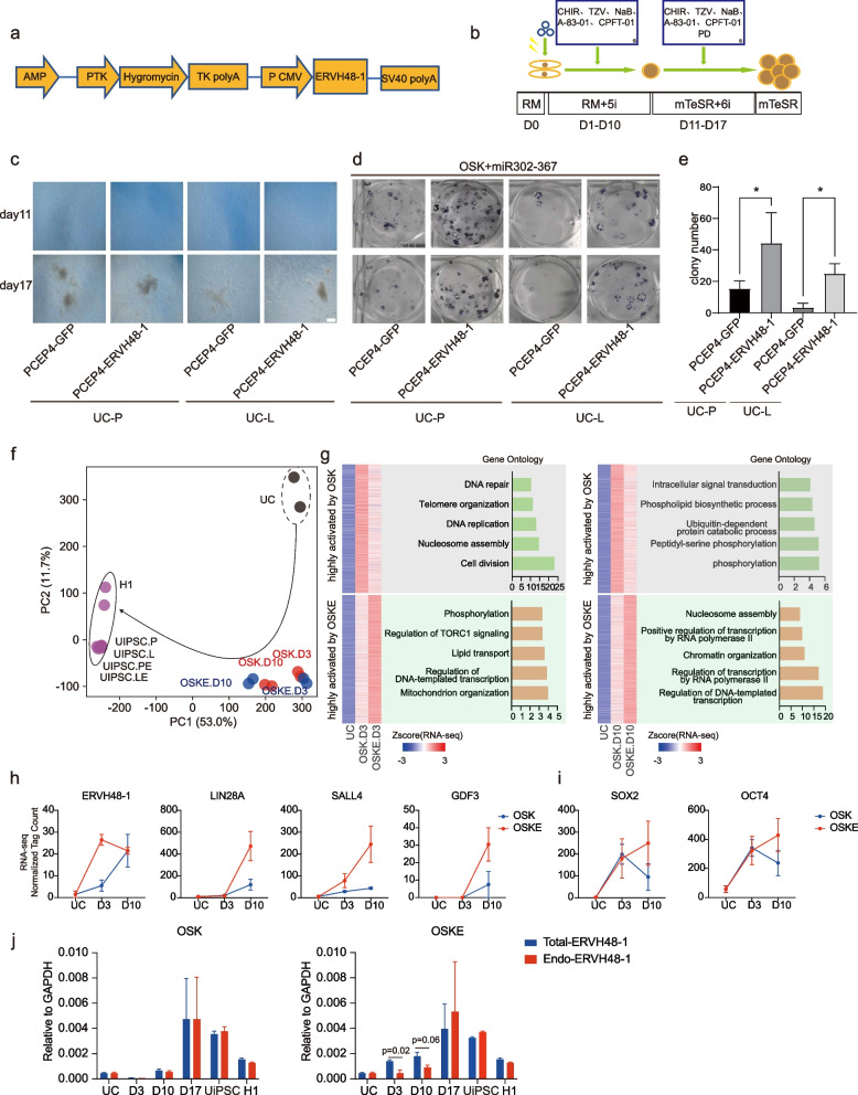
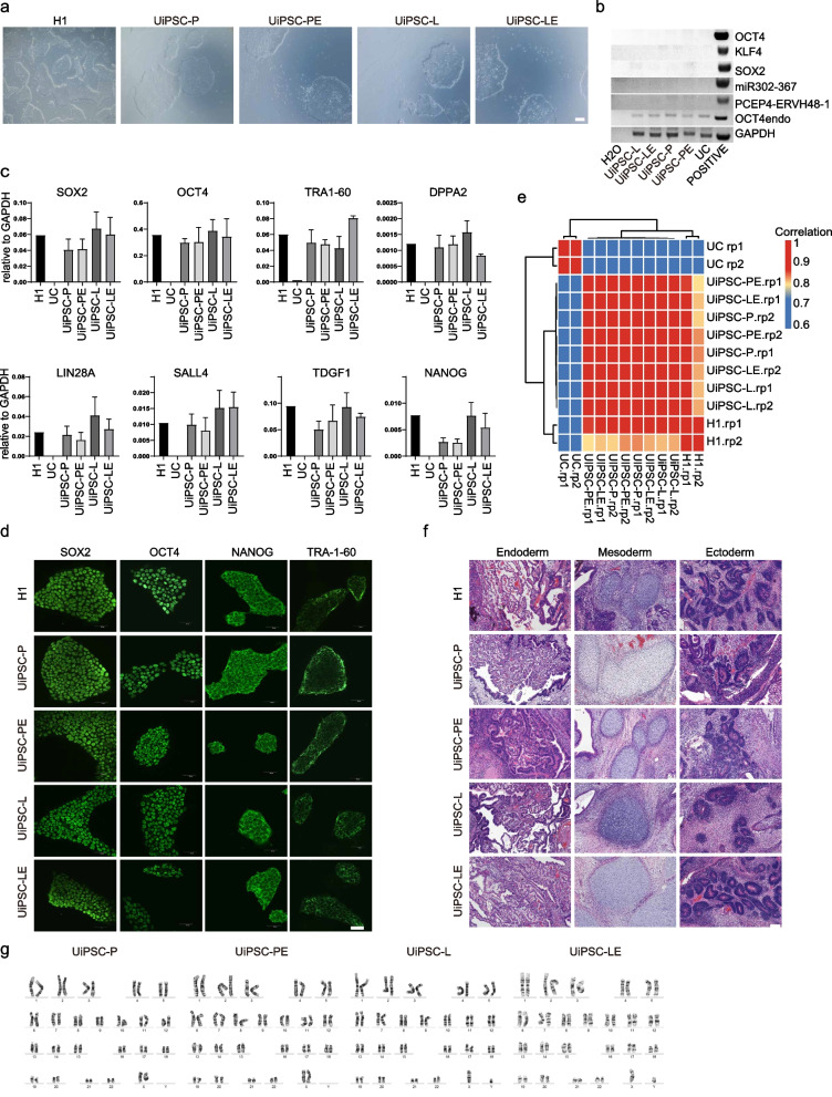
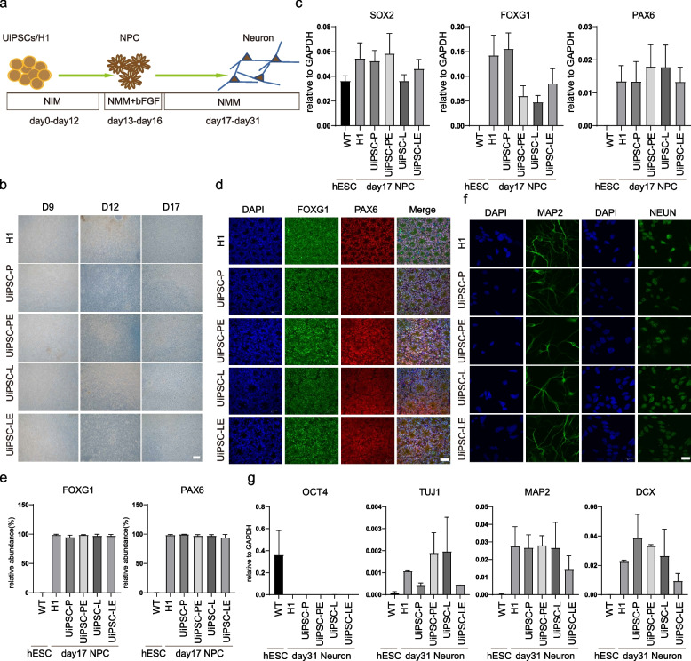
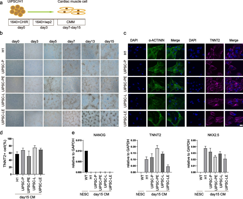
Endogenous retroviruses (ERVs), once thought to be mere remnants of ancient viral integrations in the mammalian genome, are now recognized for their critical roles in various physiological processes, including embryonic development, innate immunity, and tumorigenesis. Their impact on host organisms is significant driver of evolutionary changes, offering insight into evolutionary mechanisms. In our study, we explored the functionality of ERVs by examining single-cell transcriptomic profiles from human embryonic stem cells and urine cells. This led to the discovery of a unique ERVH48-1 expression pattern between these cell types. Additionally, somatic cell reprogramming efficacy was enhanced when ERVH48-1 was overexpressed in a urine cell-reprogramming system. Induced pluripotent stem cells (iPSCs) generated with ERVH48-1 overexpression recapitulated the traits of those produced by traditional reprogramming approaches, and the resulting iPSCs demonstrated the capability to differentiate into all three germ layers in vitro. Our research elucidated the role of ERVs in somatic cell reprogramming.

Keywords: ERVH48-1; Endogenous retroviruses; Human embryonic stem cells; Induced pluripotent stem cells; Urine cell integration-free reprogramming system.


Conflict of interest statement

Duanqing Pei is a member of the Editorial Board for Cell Regeneration. He was not involved in the journal’s review of, or decisions related to, this manuscript.

Figures

Fig. 1
Fig. 1
Transcriptome sequencing analysis reveals the expression of ERVH48-1 in hPSCs. a Heat maps of RNA-seq analysis results for UCs and H1 (hPSCs) showing 499 highly expressed genes in H1 and 425 in UCs. b Gene ontology analysis of UCs and H1. c Cluster analysis diagram of UCs and H1. d Genes with high expression in UCs and H1 were identified from the cluster, revealing ERV series expression in H1 but not in UCs. e Violin plot of the screened genes
Fig. 2
Fig. 2
ERVH48-1 overexpression promotes the reprogramming of UCs to iPSCs. a Construction map of the ERVH48-1 overexpression vector: pCEP4-ERVH48-1. b UiPSCs acquisition protocol: the reprogramming-related plasmids were electroporated into UCs, RM plus 5i medium was used during reprogramming, RM plus 6i medium was used during proliferation, and mTeSR medium was used during purification. c Morphological changes of cells at different time points during the acquisition of UiPSCs from UCs of two healthy volunteers (scale: 500 μm). d AP staining of UiPSCs from two healthy volunteers. e Number of AP + clones of UiPSCs in each group. Data were from three independent experiments and are shown as the mean ± SEM. *P < 0.05, unmatched two-tailed T-test. f PCA analysis of the bulk RNA-seq data during reprogramming of UCs into iPSCs with or without ERVH48-1. OSK: reprograming with pCEP4-OSK + miR302-367 + GFP; OSKE: reprogramming with pCEP4-OSK + miR302-367 + ERVH48-1. D3: day 3; D10: day 10. g Heatmap showing differentially expressed genes among UC, OSK, and OSKE groups at D3 and D10. Related gene ontology analysis is on the right of the heatmap. h Selected marker genes highly activated by OSKE. i Expression levels of pluripotent transcription factors SOX2 and OCT4 in OSK and OSKE conditions. j Expression levels of endogenous ERVH48-1 (Endo-ERVH48-1) and Total-ERVH48-1 in OSK, OSKE conditions and UCs, iPSCs, and H1
Fig. 3
Fig. 3
Quality testing of ERVH48-1-generated UiPSCs. a The morphology of UiPSCs resembles that of hPSC (scale: 500 μm). b Genomic DNA polymerase chain reaction (PCR) assay of UiPSCs cell line to analyze exogenous reprogramming factor integration. c qPCR results for pluripotent markers in UiPSCs cell lines. Data were from three independent experiments and are shown as the mean ± SEM. d Immunofluorescence images of pluripotent markers (OCT4, SOX2, NANOG, and TRA-1–60) in UiPSCs cell lines (scale: 50 μm). e Correlation coefficient matrix between UiPSCs cell line, hPSC, and UCs. f Teratoma detection in UiPSCs and hPSCs. HE staining was performed 7 weeks after subcutaneous injection, with histology consistent with endoderm, mesoderm, or ectoderm. (Scale: 100 μm). g Karyotypic analysis of these iPSC clones
Fig. 4
Fig. 4
UiPSCs can differentiate into neural cells in vitro. a Schematic diagram of the neural differentiation process. b Cell morphology at different time points during neuronal differentiation of the cell lines. c Relative mRNA expression levels of neural precursor markers (PAX6 and FOXG1) and pluripotency markers (SOX2) in cells on day 17 of differentiation. Data were from three independent experiments and are shown as the mean ± SEM. d The protein expression of neural precursor markers PAX6 and FOXG1 was detected by immunofluorescence assay on day 17 of differentiation. Nuclei were stained with DAPI (scale: 50 μm). e Flow cytometry performed on day 17 of neuronal differentiation, and the proportion of PAX6+ and FOXG1+ cells was recorded. Data were from three independent experiments and are shown as the mean ± SEM. f Immunofluorescence assay detected the expression of neuronal markers MAP2 and NEUN in the cells on day 31 of differentiation. Nuclei was stained with DAPI (scale: 50 μm). g Relative mRNA expression levels of the pluripotency markers (OCT4) and neuronal markers (TUJ1, MAP2, DCX) in cells on day 31 of differentiation. Data were from three independent experiments and are shown as the mean ± SEM
Fig. 5
Fig. 5
UiPSCs can differentiate into cardiomyocytes in vitro. a Schematic diagram of the myocardial differentiation process. b Cell morphology at different time points during myocardial differentiation from UiPSCs and hPSCs. c Immunofluorescence assay detected the expression of TNNT2 and α-actin in differentiated cardiomyocytes. Nuclei was stained with DAPI (scale: 20 μm). d Proportion of TNNT2 + cells after 15 days of cardiomyocyte differentiation was measured by flow cytometry. e qPCR detection of the expression of pluripotency marker NANOG and cardiomyocyte markers NKX2-5, TNNT2. Data were from three independent experiments and are shown as the mean ± SEM
Fig. 6
Fig. 6
UiPSCs can differentiate into lung progenitor cells in vitro. a Schematic diagram of lung progenitor cell differentiation. b Cell morphology at different time points during cell line differentiation into lung progenitor cells (scale: 200 μm). c Detection of lung progenitor cell markers (NKX2.1 and EPCAM) by immunofluorescence assay on day 15 after differentiation. Nuclear staining with DAPI (scale: 100 μm). d Flow cytometry detection of pulmonary progenitor cell markers (CD47 + &CD26- and EPCAM) on day 15 of differentiation. e Detection of lung precursor cell markers (NKX2.1 and EPCAM) in cells on day 15 of differentiation by qPCR

References

    1. Abedini A, Zhu YO, Chatterjee S, Halasz G, Devalaraja-Narashimha K, Shrestha R, Balzer MS, Park J, Zhou T, Ma Z, et al. Urinary Single-Cell Profiling Captures the Cellular Diversity of the Kidney. J Am Soc Nephrol. 2021;32:614–27. 10.1681/ASN.2020050757. 10.1681/ASN.2020050757 - DOI - PMC - PubMed
    1. Chen B, Xu K, Zhang Y, Xu P, Li C, Liu J, Xu Y. LncRNA ERVH48–1 Contributes to the Drug Resistance of Prostate Cancer and Proliferation through Sponging of miR-4784 to the Activation of the Wnt/beta-Catenin Pathway. Cancers (Basel). 2023;15(6):1902. 10.3390/cancers15061902. 10.3390/cancers15061902 - DOI - PMC - PubMed
    1. Chen J, Liu H, Liu J, Qi J, Wei B, Yang J, Liang H, Chen Y, Chen J, Wu Y, et al. H3K9 methylation is a barrier during somatic cell reprogramming into iPSCs. Nat Genet. 2013;45:34–42. 10.1038/ng.2491. 10.1038/ng.2491 - DOI - PubMed
    1. Chen M, Jia L, Zheng X, Han M, Li L, Zhang L. Ancient Human Endogenous Retroviruses Contribute to Genetic Evolution and Regulate Cancer Cell Type-Specific Gene Expression. Cancer Res. 2022;82:3457–73. 10.1158/0008-5472.CAN-22-0290. 10.1158/0008-5472.CAN-22-0290 - DOI - PubMed
    1. Chen M, Huang X, Wang C, Wang S, Jia L, Li L. Endogenous retroviral solo-LTRs in human genome. Front Genet. 2024;15:1358078. 10.3389/fgene.2024.1358078. 10.3389/fgene.2024.1358078 - DOI - PMC - PubMed

LinkOut - more resources