. 2024 Oct 7;34(19):4464-4475.e9.
doi: 10.1016/j.cub.2024.08.021. Epub 2024 Sep 12.

Protofilament-specific nanopatterns of tubulin post-translational modifications regulate the mechanics of ciliary beating

Affiliations

Protofilament-specific nanopatterns of tubulin post-translational modifications regulate the mechanics of ciliary beating

Gonzalo Alvarez Viar et al. Curr Biol. .

Abstract

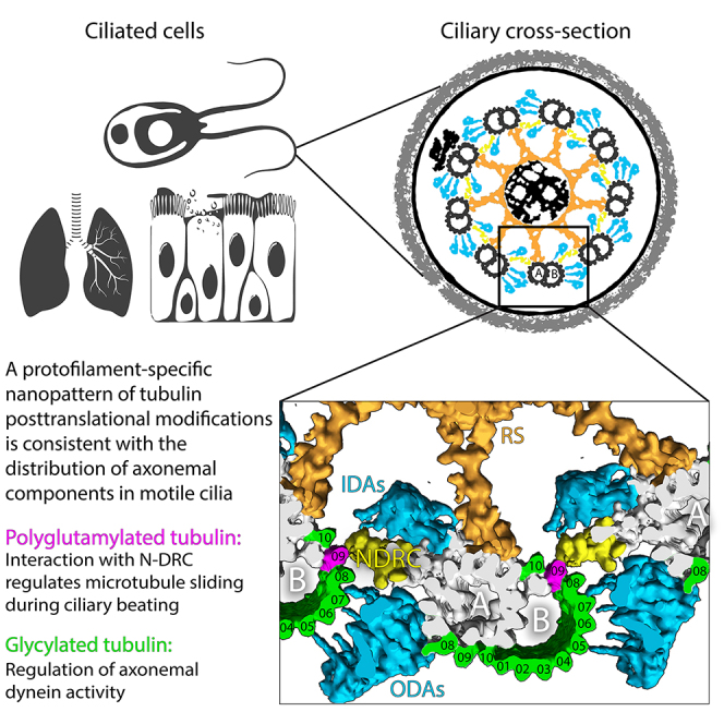
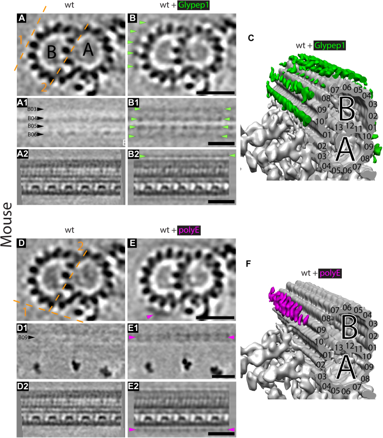
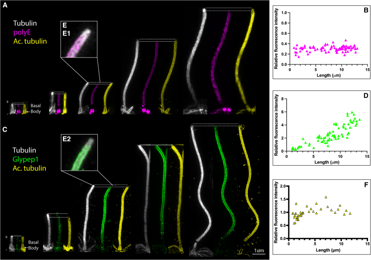
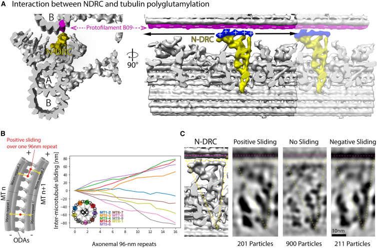
Controlling ciliary beating is essential for motility and signaling in eukaryotes. This process relies on the regulation of various axonemal proteins that assemble in stereotyped patterns onto individual microtubules of the ciliary structure. Additionally, each axonemal protein interacts exclusively with determined tubulin protofilaments of the neighboring microtubule to carry out its function. While it is known that tubulin post-translational modifications (PTMs) are important for proper ciliary motility, the mode and extent to which they contribute to these interactions remain poorly understood. Currently, the prevailing understanding is that PTMs can confer functional specialization at the level of individual microtubules. However, this paradigm falls short of explaining how the tubulin code can manage the complexity of the axonemal structure where functional interactions happen in defined patterns at the sub-microtubular scale. Here, we combine immuno-cryo-electron tomography (cryo-ET), expansion microscopy, and mutant analysis to show that, in motile cilia, tubulin glycylation and polyglutamylation form mutually exclusive protofilament-specific nanopatterns at a sub-microtubular scale. These nanopatterns are consistent with the distributions of axonemal dyneins and nexin-dynein regulatory complexes, respectively, and are indispensable for their regulation during ciliary beating. Our findings offer a new paradigm for understanding how different tubulin PTMs, such as glycylation, glutamylation, acetylation, tyrosination, and detyrosination, can coexist within the ciliary structure and specialize individual protofilaments for the regulation of diverse protein complexes. The identification of a ciliary tubulin nanocode by cryo-ET suggests the need for high-resolution studies to better understand the molecular role of PTMs in other cellular compartments beyond the cilium.

Keywords: Chlamydomonas; U-ExM; cilia motility; cryo-ET; microtubule protofilaments; molecular nanopatterns; motile cilia; tubulin code; tubulin glycylation; tubulin polyglutamylation; tubulin posttranslational modification.


Conflict of interest statement

Declaration of interests The authors declare no competing interests.

Figures

None
Graphical abstract
Figure 1
Figure 1
Localization of tubulin glycylation and polyglutamylation in the Chlamydomonas axoneme (A) Cartoon representing the ciliary cross-section and a close-up view of the 96-nm repeat showing the spatially restricted interactions between axonemal components and different protofilaments of the adjacent doublet. (B–I) Slices through 3D electron density models of the Chlamydomonas axonemal 96-nm repeat generated by sub-tomogram averaging. Dashed lines 1 and 2 in (B) depict the planes at which the different longitudinal sections were made. (B) Wild-type axoneme. (C) Wild-type axoneme decorated with Glypep1 antibodies. Note the presence of extra densities, indicated by green arrowheads, over the microtubule doublet surface recapitulating the tubulin dimer periodicity, indicating the presence of the antibody labeling and the position of glycylated tubulin.(D) ttll3− axoneme. (E) ttll3− axoneme incubated with Glypep1 antibodies. Note the absence of any labeling. (F) Wild-type axoneme. (G) Wild-type axoneme decorated with polyE antibodies. Note the presence of extra densities, indicated by magenta arrowheads, present over protofilament B09. (H) tpg1 axoneme. (I) tpg1 axonemes incubated with polyE antibodies. Note the reduction in the labeling signal compared with (G). (J) Electron density model of the ida5 96-nm repeat decorated with Glypep1 antibodies (green). (K) Electron density model of the ida5 96-nm repeat decorated with polyE antibodies (magenta). (A) and (B) labels indicate the different tubules in the microtubule doublet. Black and white arrowhead labels indicate different protofilaments. Scale bars, 20 nm. More information about the labeling strategy can be found in Figure S1. More information about the structure of the mutants can be found in Figure S2. More information about the labeling of protofilament B09 can be found in Figure S3. More information about the complementarity of glycylation and polyglutamylation patterns can be found in Figure S4. More information about the presence of polyglutamylation along protofilament B09 can be found in Figure S5. See also Table S1 for information about the Electron Microscopy Database (EMD) codes.
Figure 2
Figure 2
Localization of tubulin glycylation and polyglutamylation in the mouse respiratory axoneme Slices through 3D electron density models of the mouse respiratory ciliary axonemal 96-nm repeat generated by sub-tomogram averaging. Dashed lines depict the planes at which the different longitudinal sections were made. (A) and (B) labels indicate the different tubules in the microtubule doublet. Black arrowhead labels indicate different protofilaments. (A) Mouse respiratory ciliary axonemes isolated from trachea tissue. (B) Mouse respiratory ciliary axonemes decorated with Glypep1 antibodies. Note the presence of extra densities, indicated by green arrowheads, over the microtubule doublet surface recapitulating the tubulin dimer periodicity. (C) Electron density model of the mouse respiratory ciliary axoneme decorated with Glypep1 antibodies (green). (D) Mouse respiratory ciliary axonemes isolated from trachea tissue. (E) Mouse respiratory ciliary axonemes decorated with polyE antibodies. Note the presence of extra densities, indicated by magenta arrowheads, over protofilament B09. (F) Electron density model of the mouse respiratory ciliary axoneme decorated with polyE antibodies (magenta). Scale bars, 20 nm. More information about the structure of the mouse respiratory ciliary axoneme can be found in Figure S6. See also Table S1 for information about the EMD codes.
Figure 3
Figure 3
Axonemal incorporation of tubulin polyglutamylation and glycylation during ciliary regrowth in Chlamydomonas Ultrastructural expansion microscopy of regrowing flagella of Chlamydomonas. (A) Triple immunostaining of regrowing Chlamydomonas axonemes against tubulin (gray), polyglutamylated tubulin (magenta), and acetylated tubulin (yellow). Note that even for very short cilia, polyglutamylation is readily incorporated. (B) Quantification of polyE labeling density over regrowing cilia normalized to polyE signal in the basal body (representative images for this quantification can be found in Figure S7). (C) Triple immunostaining of regrowing Chlamydomonas axonemes against tubulin (gray), glycylated tubulin (green), and acetylated tubulin (yellow). Note that for short cilia, glycylation signal is sparse while its density increases for long cilia. (D) Quantification of Glypep1 labeling density over regrowing cilia normalized to centrin signal in the basal body (representative images for this quantification can be found in Figure S8). (E1 and E2) Blow-up from a single ciliary tip showing the overhang of the tubulin signal with respect to the respective tubulin PTMs. (F) Quantification of acetylated tubulin labeling density over regrowing cilia normalized to acetylated tubulin signal in the basal body. Scale bar, 1 μm.
Figure 4
Figure 4
Tubulin glycylation is required for normal cell swimming behavior (A) 3D electron density model of the Chlamydomonas 96-nm repeat showing the interaction between ODAs and IDAs (turquoise) with glycylated tubulin (green). (B) Phototactic response (negative) of wild-type, ttll3::BSD, and ttll3::BSD TTLL3-PAR cells after 4 min of illumination with green light. Note that the pattern of phototaxis differs between wild type and ttll3::BSD and that it is recovered in the rescue strain. (C) Violin plots of swimming speed distributions in wild-type, ttll3::BSD, and ttll3::BSD TTLL3-PAR cells. (D) Violin plots of beating frequency distributions in wild-type, ttll3::BSD, and ttll3::BSD TTLL3-PAR cells. (E) Violin plots of mean total displacement per beat cycle distributions in wild-type, ttll3::BSD, and ttll3::BSD TTLL3-PAR cells. p values were calculated in python using a t test (scipy.stats.ttest_ind). More information about the characterization of the phenotype can be found in Figure S9. See also Data S1.
Figure 5
Figure 5
Inter-microtubule doublet sliding quantification and characterization of N-DRC structure during ciliary bending (A) Electron density model of the interaction between the N-DRC (yellow) and protofilament B09 (magenta) in wild-type Chlamydomonas cells. (B) Measurement of the inter-microtubule sliding distributions in a bent axoneme. The cartoon on the left illustrates the relationship between microtubule sliding and bending (adapted from www.macmillanhighered.com). (C) Slices through sub-tomogram average models of the N-DRC subjected to different sliding conditions showing that the N-DRC does not tilt or stretch at macromolecular level during ciliary bending in vitro. See also Table S1 for information about the EMD codes.

References

    1. Afzelius B.A. A Human Syndrome Caused by Immotile Cilia. Science. 1976;193:317–319. doi: 10.1126/science.1084576. - DOI - PubMed
    1. Badano J.L., Mitsuma N., Beales P.L., Katsanis N. The Ciliopathies: An Emerging Class of Human Genetic Disorders. Annu. Rev. Genomics Hum. Genet. 2006;7:125–148. doi: 10.1146/annurev.genom.7.080505.115610. - DOI - PubMed
    1. Waters A.M., Beales P.L. Ciliopathies: an expanding disease spectrum. Pediatr. Nephrol. 2011;26:1039–1056. doi: 10.1007/s00467-010-1731-7. - DOI - PMC - PubMed
    1. Mitchison H.M., Valente E.M. Motile and non-motile cilia in human pathology: from function to phenotypes. J. Pathol. 2017;241:294–309. doi: 10.1002/path.4843. - DOI - PubMed
    1. Goodenough U.W., Heuser J.E. Substructure of Inner Dynein Arms, Radial Spokes, and the Central Pair/Projection Complex of Cilia and Flagella. J. Cell Biol. 1985;100:2008–2018. doi: 10.1083/jcb.100.6.2008. - DOI - PMC - PubMed

MeSH terms

LinkOut - more resources