. 2024 Oct 3;31(10):1513-1523.e7.
doi: 10.1016/j.stem.2024.08.009. Epub 2024 Sep 12.

Human pluripotent stem cell-derived organoids repair damaged bowel in vivo

Affiliations

Human pluripotent stem cell-derived organoids repair damaged bowel in vivo

Holly M Poling et al. Cell Stem Cell. .

Abstract

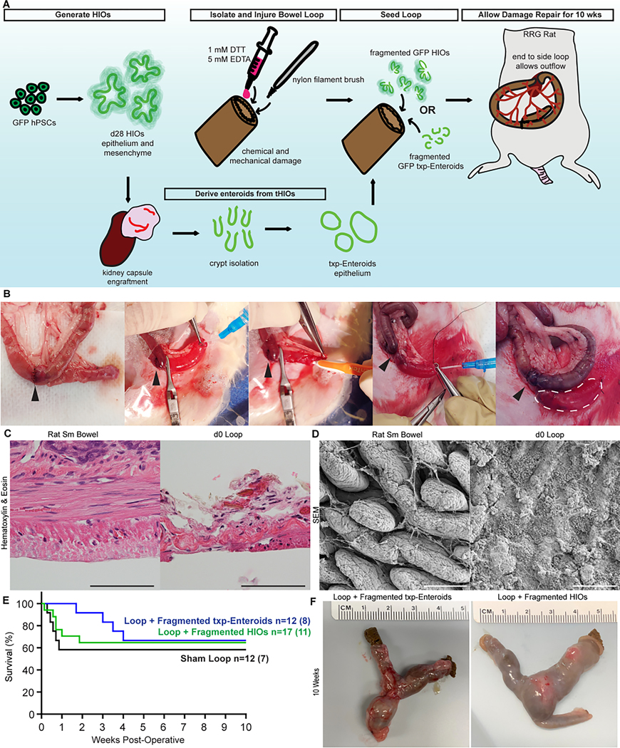
The fundamental goal of tissue engineering is to functionally restore or improve damaged tissues or organs. Here we address this in the small bowel using an in vivo xenograft preclinical acute damage model. We investigated the therapeutic capacity of human intestinal organoids (HIOs), which are generated from human pluripotent stem cells (hPSCs), to repair damaged small bowel. We hypothesized that the HIO's cellular complexity would allow it to sustain transmural engraftment. To test this, we developed a rodent injury model where, through luminal delivery, we demonstrated that fragmented HIOs engraft, proliferate, and persist throughout the bowel following repair. Not only was restitution of the mucosal layer observed, but significant incorporation was also observed in the muscularis and vascular endothelium. Further analysis characterized sustained cell type presence within the regenerated regions, retention of proximal regionalization, and the neo-epithelia's function. These findings demonstrate the therapeutic importance of mesenchyme for intestinal injury repair.

Keywords: cell therapy; enteroid; human intestinal organoid; intestine; tissue regeneration.


Conflict of interest statement

Declaration of interests CCHMC has a patent application in process related to the work in this study.

Figures

Figure 1.
Figure 1.. Experimental Design and Mucosectomy Surgical Injury Model.
(A) Experimental design schematic. (B) Surgical imagery of loop creation, damage, and application of cell therapy product. Arrowhead indicates anastomosis. White dashed line outlines loop. (C) Histology of healthy and injured rat small bowel. (D) Topography of healthy and injured rat small bowel. (E) Kaplan-Meier curve associated with surgeries. (F) Harvest images of damaged loops reseeded with txp-Enteroids and HIOs after 10 weeks. All scale bars = 100 μm.
Figure 2.
Figure 2.. Organoid Fragmentation was Essential for Expansion during Regeneration.
(A) Representative histology of a Loop + Whole HIOs and serial section stained for human cells (KU80, brown). (B) Brightfield and live-GFP fluorescence imagery of HIOs and txp-Enteroids before and after fragmentation. (C) Histograms of HIO and txp-Enteroid fragment sizes. (D) Graph of cell number in HIOs and txp-Enteroids represented as mean ± SD. (E) Brightfield and live-GFP fluorescence image of a harvested Loop + Fragmented HIOs after 10 weeks; dashed line indicates tissue edge. (F) Quantification of live-GFP surface area in Loop + Fragmented HIOs samples. (G) Representative section stained for human cells (KU80, brown) in a live-GFP+ region in a Loop + Fragmented HIO. Boxed inset depicts margin/boundary of human (KU80, brown) and rat tissue. (H) Quantification of KU80+ cells as a percentage of cells in each 20x field of view for areas with positive human cellular incorporation in Loop + Fragmented HIOs samples. (I) Brightfield and live-GFP fluorescence image of a harvested Loop + Fragmented txp-Enteroids after 10 weeks; dashed line indicates tissue edge. (J) Quantification of live-GFP surface area in Loop + Fragmented txp-Enteroids samples. (K) Representative section stained for human cells (KU80, brown) in a live-GFP+ region in a Loop + Fragmented txp-Enteroids. (L) Quantification of KU80+ cells as a percentage of cells in each 20x field of view for areas with positive human cellular incorporation in Loop + Fragmented txp-Enteroids samples. All scale bars = 100 μm. For F vs. J, p=0.0023 and for H vs. L, p<0.0001 from Mann-Whitney Tests.
Fig. 3.
Fig. 3.. Fragmented HIOs contribute to tissue regeneration of damaged bowel in vivo after 10 weeks.
(A) Tile scan of a Loop + Fragmented HIOs stained for human cells (KU80, brown); scale bar = 0.5 cm. Higher magnification images of KU80+ cells within the mucosa and muscularis from tile scan; scale bar = 50 μm. (B) Representative images of Sham Loop and Loop + Fragmented HIOs stained for a proliferation marker (MKI67, pink), epithelium (CDH1, white) and human cells (GFP, green). (C) Representative images of Sham Loop and Loop + Fragmented HIOs stained for a surrogate marker of stem cell activity and the intestinal crypt (OLFM4, pink) and epithelium (CDH1, green). (D) Representative images of Sham Loop and Loop + Fragmented HIOs stained for human cells (KU80, brown) and goblet cells (alcian blue for mucin, blue). (E) Representative images of Sham Loop and Loop + Fragmented HIOs stained for human cells (GFP, green) and Paneth cells (DEFA5, pink). (F) Representative images of Sham Loop and Loop + Fragmented HIOs stained for human cells (GFP, green), and enteroendocrine cells (CHGA, white). (G) Representative images of Sham Loop and Loop + Fragmented HIOs stained for human cells (GFP, green), and smooth muscle (ACTA2, pink). (H) Representative images of Sham Loop and Loop + Fragmented HIOs stained for a pan-neuronal marker (TUBB3, brown). Arrow heads indicate neuronal bundles. (I) Representative images of Sham Loop and Loop + Fragmented HIOs stained for human cells (GFP, green), blood vessels (VWF, pink), and human blood vessels (hCD31, white). Single channel insets are within the white dashed box. (B-I) Scale bars = 50 μm.
Fig. 4.
Fig. 4.. Neo-epithelia of reseeded loops are responsive to chemical stimuli after 10 weeks.
(A) Representative merged brightfield and live-GFP images of healthy rat small bowel (proximal to the loop), Sham Loop, and Loop + Fragmented HIOs mounted in an Ussing assay slider; dashed circle outlines slider opening. (B) Representative time course of healthy rat small bowel Isc measured in Ussing Chamber experiments. (C) Representative time course of Sham Loop Isc measured in Ussing Chamber experiments. (D) Representative time course of Loop + Fragmented HIOs Isc measured in Ussing Chamber experiments. (E) Graphs of calculated changes in Isc in response to 10 μM Forskolin, 100 μM IBMX, and 100 μM Bumetanide across sample groups. (F) Graph of baseline TEER across sample groups. (G) Graph of FITC-dextran permeability across sample groups. (H) Calculated FITC flux from graph in G. Bar graphs are represented as mean ± SD. Kruskal-Wallis test p-values were not significant between groups in E-H.

References

    1. Singh A, Poling HM, Spence JR, Wells JM, and Helmrath MA (2020). Gastrointestinal organoids: a next-generation tool for modeling human development. Am J Physiol Gastrointest Liver Physiol 319, G375–G381. 10.1152/ajpgi.00199.2020. - DOI - PMC - PubMed
    1. Sato T, and Clevers H. (2013). Growing self-organizing mini-guts from a single intestinal stem cell: mechanism and applications. Science 340, 1190–1194. 10.1126/science.1234852. - DOI - PubMed
    1. Yui S, Nakamura T, Sato T, Nemoto Y, Mizutani T, Zheng X, Ichinose S, Nagaishi T, Okamoto R, Tsuchiya K, et al. (2012). Functional engraftment of colon epithelium expanded in vitro from a single adult Lgr5(+) stem cell. Nat Med 18, 618–623. 10.1038/nm.2695. - DOI - PubMed
    1. Watanabe S, Kobayashi S, Ogasawara N, Okamoto R, Nakamura T, Watanabe M, Jensen KB, and Yui S. (2022). Transplantation of intestinal organoids into a mouse model of colitis. Nat Protoc 17, 649–671. 10.1038/s41596-021-00658-3. - DOI - PubMed
    1. Avansino JR, Chen DC, Hoagland VD, Woolman JD, and Stelzner M. (2006). Orthotopic transplantation of intestinal mucosal organoids in rodents. Surgery 140, 423–434. 10.1016/j.surg.2006.03.012. - DOI - PubMed

LinkOut - more resources