

Subcutaneous edema segmentation on abdominal CT using multi-class labels and iterative annotation
- PMID: 39271574
- PMCID: PMC11757642
- DOI: 10.1007/s11548-024-03262-4
Subcutaneous edema segmentation on abdominal CT using multi-class labels and iterative annotation
Abstract
Purpose: Anasarca is a condition that results from organ dysfunctions, such as heart, kidney, or liver failure, characterized by the presence of edema throughout the body. The quantification of accumulated edema may have potential clinical benefits. This work focuses on accurately estimating the amount of edema non-invasively using abdominal CT scans, with minimal false positives. However, edema segmentation is challenging due to the complex appearance of edema and the lack of manually annotated volumes.
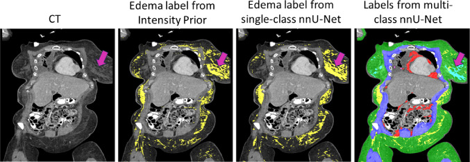
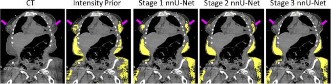
Methods: We propose a weakly supervised approach for edema segmentation using initial edema labels from the current state-of-the-art method for edema segmentation (Intensity Prior), along with labels of surrounding tissues as anatomical priors. A multi-class 3D nnU-Net was employed as the segmentation network, and training was performed using an iterative annotation workflow.
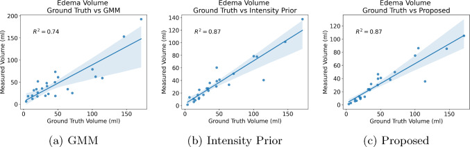
Results: We evaluated segmentation accuracy on a test set of 25 patients with edema. The average Dice Similarity Coefficient of the proposed method was similar to Intensity Prior (61.5% vs. 61.7%; ). However, the proposed method reduced the average False Positive Rate significantly, from 1.8% to 1.1% ( ). Edema volumes computed using automated segmentation had a strong correlation with manual annotation ( ).
Conclusion: Weakly supervised learning using 3D multi-class labels and iterative annotation is an efficient way to perform high-quality edema segmentation with minimal false positives. Automated edema segmentation can produce edema volume estimates that are highly correlated with manual annotation. The proposed approach is promising for clinical applications to monitor anasarca using estimated edema volumes.
Keywords: Anasarca; Edema segmentation; Iterative annotation; Weakly supervised learning; nnU-Net.
© 2024. This is a U.S. Government work and not under copyright protection in the US; foreign copyright protection may apply.
Conflict of interest statement
Declarations. Conflict of interest: RMS receives royalties from iCAD, Philips, PingAn, ScanMed, MGB, and Translation Holdings. His lab received research support from PingAn. The authors have no additional Conflict of interest to declare. Ethics approval: The study was approved by the IRB of the National Institutes of Health and was performed in accordance with the ethical standards as laid down in the 1964 Declaration of Helsinki and its later amendments. Consent to participate: The need for written informed consent was waived by the IRB.
Figures


Similar articles
-
Weakly supervised learning for subcutaneous edema segmentation of abdominal CT using pseudo-labels and multi-stage nnU-Nets.Proc SPIE Int Soc Opt Eng. 2024 Feb;12927:1292738. doi: 10.1117/12.3008793. Epub 2024 Apr 3. Proc SPIE Int Soc Opt Eng. 2024. PMID: 39371589 Free PMC article.
-
Cooperative AI training for cardiothoracic segmentation in computed tomography: An iterative multi-center annotation approach.Eur J Radiol. 2024 Jul;176:111534. doi: 10.1016/j.ejrad.2024.111534. Epub 2024 May 25. Eur J Radiol. 2024. PMID: 38820951
-
Efficient contour-based annotation by iterative deep learning for organ segmentation from volumetric medical images.Int J Comput Assist Radiol Surg. 2023 Feb;18(2):379-394. doi: 10.1007/s11548-022-02730-z. Epub 2022 Sep 1. Int J Comput Assist Radiol Surg. 2023. PMID: 36048319 Free PMC article.
-
Abdominal multi-organ segmentation from CT images using conditional shape-location and unsupervised intensity priors.Med Image Anal. 2015 Dec;26(1):1-18. doi: 10.1016/j.media.2015.06.009. Epub 2015 Jul 4. Med Image Anal. 2015. PMID: 26277022 Free PMC article.
-
A unified approach to medical image segmentation by leveraging mixed supervision and self and transfer learning (MIST).Comput Med Imaging Graph. 2025 Jun;122:102517. doi: 10.1016/j.compmedimag.2025.102517. Epub 2025 Mar 5. Comput Med Imaging Graph. 2025. PMID: 40088573
Cited by
-
Progress in fully automated abdominal CT interpretation-an update over the past decade.Abdom Radiol (NY). 2025 Jul 8. doi: 10.1007/s00261-025-05094-5. Online ahead of print. Abdom Radiol (NY). 2025. PMID: 40627132 Review.
References
-
- Kattula S, Avula A, Baradhi K (2021) Anasarca, StatPearls. StatPearls Publishing, Treasure Island (FL) - PubMed
-
- Having K, Bullock S (2011) Fetal anasarca. J Diagn Med Sonogr 27(1):19–25
-
- Bobkova I, Chebotareva N, Kozlovskaya L, Shilov E (2016) Edema in renal diseases-current view on pathogenesis. Nephrol Point Care 2(1):5000204
-
- Dierckx R, Haine SE, Vrints CJ, Paelinck BP (2008) Young adult with congenital heart disease presenting with anasarca. Circulation 118(12):1304–1305 - PubMed
MeSH terms
Grants and funding
LinkOut - more resources
Full Text Sources
Medical
