. 2024 Sep 13;15(1):8017.
doi: 10.1038/s41467-024-51971-w.

HIV-1 adapts to lost IP6 coordination through second-site mutations that restore conical capsid assembly

Affiliations

HIV-1 adapts to lost IP6 coordination through second-site mutations that restore conical capsid assembly

Alex Kleinpeter et al. Nat Commun. .

Abstract

The HIV-1 capsid is composed of capsid (CA) protein hexamers and pentamers (capsomers) that contain a central pore hypothesised to regulate capsid assembly and facilitate nucleotide import early during post-infection. These pore functions are mediated by two positively charged rings created by CA Arg-18 (R18) and Lys-25 (K25). Here we describe the forced evolution of viruses containing mutations in R18 and K25. Whilst R18 mutants fail to replicate, K25A viruses acquire compensating mutations that restore nearly wild-type replication fitness. These compensating mutations, which rescue reverse transcription and infection without reintroducing lost pore charges, map to three adaptation hot-spots located within and between capsomers. The second-site suppressor mutations act by restoring the formation of pentamers lost upon K25 mutation, enabling closed conical capsid assembly both in vitro and inside virions. These results indicate that there is no intrinsic requirement for K25 in either nucleotide import or capsid assembly. We propose that whilst HIV-1 must maintain a precise hexamer:pentamer equilibrium for proper capsid assembly, compensatory mutations can tune this equilibrium to restore fitness lost by mutation of the central pore.


Conflict of interest statement

The authors declare no competing interests.

Figures
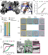
Fig. 1
Fig. 1. Mutation of R18G and K25A profoundly reduces infection but differentially impacts capsid formation.
A Cross-section of a CA hexamer showing two IP6 molecules bound within the pore by the charged rings at R18 and K25 (based on 6R6Q). Side panel shows a close-up view. B Titration of WT and mutant HIV-1 VSV-G-pseudotyped virus on HEK293T cells with infectivity quantified as the proportion of infected cells (area of monolayer). Error bars depict mean ± s.e.m. from at least three independent experiments (N = 3). C Cryo-ET analysis of the indicated HIV-1 mutants. Tilt series were collected and reconstructions performed to assess capsid morphology. A total of 51 WT, 38 K25A and 43 R18G particles were analyzed and classified into the indicated categories. Example sliced tomograms belonging to each category are shown together with the data for each virion. Scale bars, 100 nm. D In vitro assembly of 900 µM CA (WT, K25A and R18G) in 6 mM IP6, measuring absorbance of reaction over time at 350 nm. Negative stain EM images of assembly reactions are shown with 4x zoomed in sections displayed above. Scalebar: 200 nm.
Fig. 2
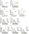
Fig. 2. K25A replication can be rescued by second-site mutations.
A, B MT4 cells were transfected with infectious molecular clones harboring mutations at either R18 (A) or K25 (B) and replication kinetics were assessed by quantifying supernatant RT activity. C Supernatants from (B) and similar repeat experiments were used to infect MT4, C8166, or SupT1 T cell lines to assess replication kinetics after acquisition of compensatory mutations. Infectious molecular clones of N21S (D), T216I (E), G208R (F), A105T (G) and G225S (H) variants were used to transfect MT4 cells to assess replication kinetics.
Fig. 3
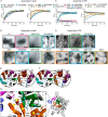
Fig. 3. Infectivity of K25A but not R18G can be rescued by second-site mutations.
A Representations of the hexamer (8CKV; left-hand side) and pentamer (8CKW; right-hand side) interactions in an HIV-1 capsid assembled in the presence of IP6. The central hexamer or pentamer is shown in green and α-helices are indicated by cylinders. An example 3-fold inter-capsomer interface is highlighted with interacting monomers in orange, blue and pink. Zoomed-in regions show the location of second-site mutations selected during passage of K25A virus, with the specific residues labelled and indicated through sphere representation of their main-chain atoms. These regions correspond to the central pore (N21 and K25), the top of the CPSF6- binding pocket (A105 and T107) and the 3-fold interface (G208 and T216). B, C Single-round infectivity of the indicated viruses as measured by the proportion of infected cells (area of monolayer). Error bars depict mean ± s.e.m. from at least three independent experiments (N = 3).
Fig. 4
Fig. 4. K25A compensating mutations restore DNA synthesis without altering nucleotide binding.
A Thermostability of the indicated crosslinked CA hexamers either alone or in the presence of different polyanions, as measured by differential scanning fluorimetry. B Change in melt temperature (ΔTm) upon polyanion addition, calculated from the data in (A). Error bars depict the s.e.m. from three independent experiments. C Binding affinities of indicated WT and mutant crosslinked CA hexamers to fluorescent ATP as measured by fluorescence polarization binding assays. Error bars depict the s.e.m. from 3 independent experiments. D Quantification of viral DNA (vDNA) produced by the indicated viruses at various time-points post-infection (in hours). Early (minus-strand strong stop; MSSS), mid (GFP) and late (second-strand transfer; SST) products are quantified using specific primers. Error bars depict mean ± s.e.m. from at least three independent experiments (N = 3).
Fig. 5
Fig. 5. K25A in vitro capsid assembly is rescued by compensating mutations.
A Assembly of 200 µM K25A (left) or K25T (right) CA variants in 2.5 mM NaCl, measuring absorbance over time at 350 nm. B Negative stain EM images of assembly reactions from (A). A, B WT data are reproduced in each pair of graphs to allow easier comparison with mutants. C Assembly of 200 µM K25A (left) or K25T (right) variants in 1.25 mM IP6. Where indicated by the asterisk, assembly reactions were performed in 400 µM CA and 10 mM IP6. D Negative stain EM images of assembly reactions from (C). Scalebar: 200 nm. EG Representations of packing interactions involving capsid pentamers (8CKW) in an HIV-1 capsid assembled in the presence of IP6. E N21-proximal residues at the interface between neighbouring monomers in the pentamer, including the key pawl and ratchet residue M39. Dotted lines from N21 highlight the relative distance and positioning of nearby residues and do not necessarily indicate non-covalent interactions. F As (E), but with S21 modelled in place of N21 and T25 instead of K25. G Interaction networks that promote packing at multiple pentamer (8CKW) interfaces. Two monomers of the same pentamer are shown in orange and green and parts of monomers from adjacent capsomers in pink and blue. A proposed allosteric network connects a linear ratchet motif (V24, K25 & M49) to a ‘TVGG’ motif and gate residue M66 that modulate hexamer or pentamer formation. The TVGG motif sits behind a three-fold interface where T216I is located. Yellow arrows indicate that interactions at each interface determine how closely monomers pack within and between pentamers.
Fig. 6
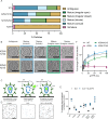
Fig. 6. Second-site mutations stabilize K25A capsids that form inside virions.
A, B Cryo-ET of the indicated HIV-1 mutants. Tilt series were collected and reconstructions performed to assess capsid morphology. A total of 51 WT, 38 K25A, 146 N21S/K25A and 67 K25A/T216I particles were analyzed and classified into the indicated categories. Example sliced tomograms belonging to each category are shown together with the data for each virion. Examples of WT and K25A tomograms can be seen in Fig. 1. Scale bars, 100 nm. C Viruses containing EGFP as a capsid content marker were bound to glass dishes and permeabilised with SLO in the presence or absence of 50 μM IP6. Images were acquired with a TIRF microscope and particles counted using Fiji. D Representative masks generated during analysis of immobilised WT virus 30 min post-SLO or control treatment ± addition of 50 µM IP6. Scale bar = 10 µm. E A single representative experiment (N = 1) where intact capsids from five 88 µm2 images were counted, typically 500–1000/image for each condition, and the fraction of intact capsids in +SLO conditions plotted as a percentage of the mean under -SLO conditions (the overall mean of the fraction of intact capsids in +SLO conditions from five images is shown as a black bar ). F TRIM5 abrogation experiment. An increasing input of the indicated RFP reporter viruses was added to cells expressing rhesus TRIM5α in the presence of a constant dose of WT GFP virus. RFP viruses with a capsid capable of binding TRIM5 will saturate the restriction factor, leading to an increase in GFP virus infection. Error bars depict mean  ± s.e.m. from at least four independent experiments (N = 4).
Fig. 7
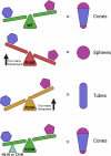
Fig. 7. Compensating mutations restore hexamer:pentamer equilibrium.
WT CA can form hexamers and pentamers but favours hexamers as more are needed to build a conical capsid. R18G shifts the equilibrium in favour of pentamers, resulting in the formation of pentamer-rich spheres in vitro. K25A shifts the equilibrium too far in favour of hexamers, resulting in the formation of hexameric tubes in vitro. Second-site compensatory mutations such as N21S or T216I stabilise pentamers, restoring WT equilibrium and thus conical capsid formation. Hexamers are blue and pentamers are purple. Note that in virions, R18G capsids are mostly irregular in their morphology whilst K25A capsids cannot be clearly assigned.

References

    1. Pornillos, O. et al. X-ray structures of the hexameric building block of the HIV capsid. Cell137, 1282–1292 (2009). 10.1016/j.cell.2009.04.063 - DOI - PMC - PubMed
    1. James, L. C. The HIV-1 Capsid: More than Just a Delivery Package. Adv. Exp. Med Biol.1215, 69–83 (2019). 10.1007/978-3-030-14741-9_4 - DOI - PubMed
    1. James, L. C. & Jacques, D. A. The human immunodeficiency virus capsid is more than just a genome package. Annu Rev. Virol.5, 209–225 (2018). 10.1146/annurev-virology-092917-043430 - DOI - PubMed
    1. Braaten, D. & Luban, J. Cyclophilin A regulates HIV-1 infectivity, as demonstrated by gene targeting in human T cells. EMBO J.20, 1300–1309 (2001). 10.1093/emboj/20.6.1300 - DOI - PMC - PubMed
    1. Dharan, A. & Campbell, E. M. Role of microtubules and microtubule-associated proteins in HIV-1 infection. J. Virol.92, e00085–18 (2018). 10.1128/JVI.00085-18 - DOI - PMC - PubMed

Publication types

Substances

LinkOut - more resources