. 2024 Sep 13;7(1):1138.
doi: 10.1038/s42003-024-06817-y.

Ligand requirements for immunoreceptor triggering

Affiliations

Ligand requirements for immunoreceptor triggering

Michael I Barton et al. Commun Biol. .

Abstract

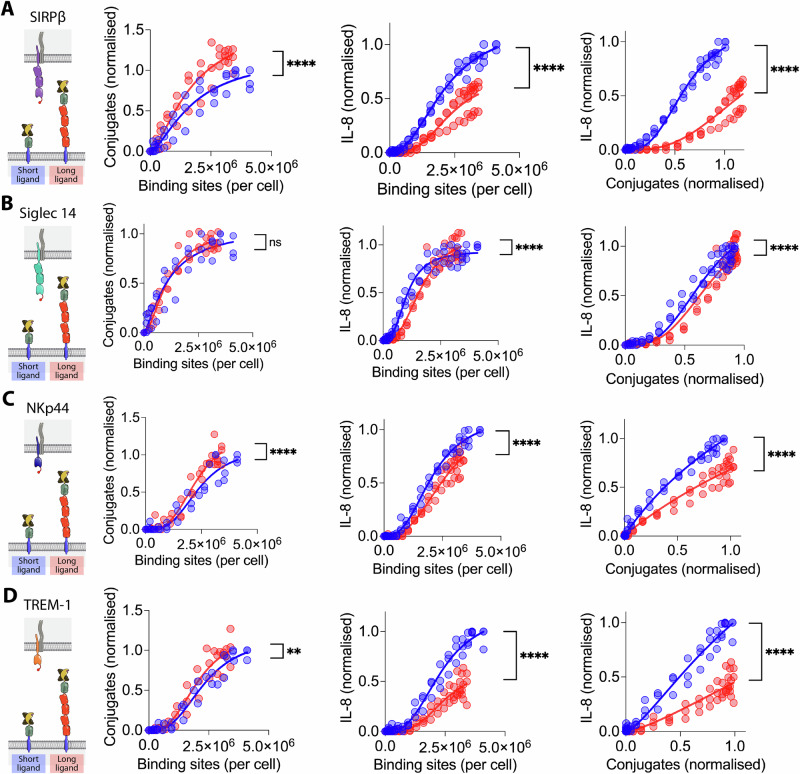
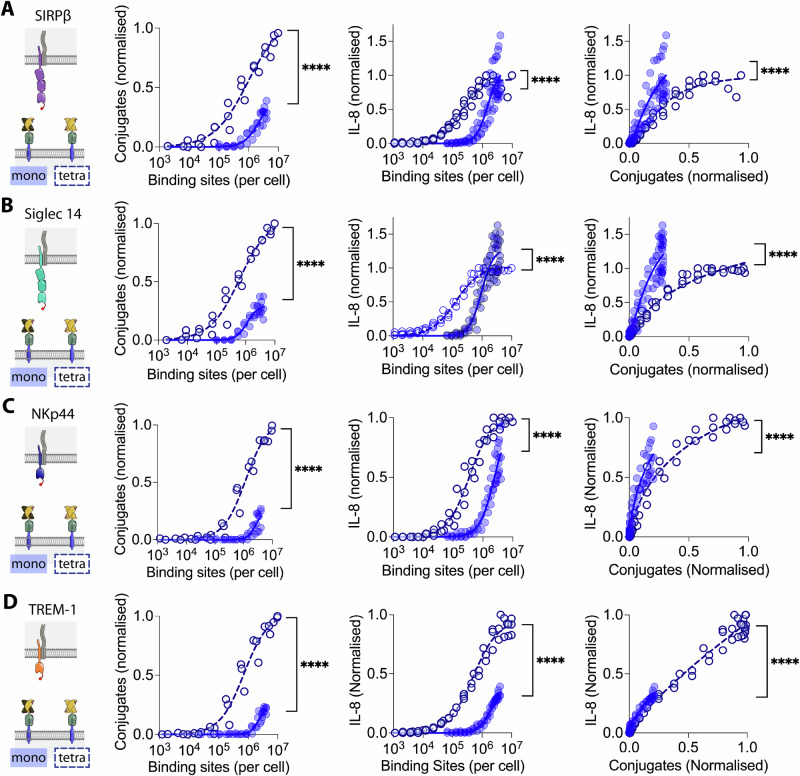
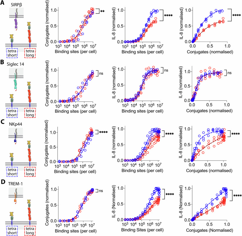
Leukocytes interact with other cells using cell surface receptors. The largest group of such receptors are non-catalytic tyrosine phosphorylated receptors (NTRs), also called immunoreceptors. NTR signalling requires phosphorylation of cytoplasmic tyrosine residues by SRC-family tyrosine kinases. How ligand binding to NTRs induces this phosphorylation, also called NTR triggering, remains controversial, with roles suggested for size-based segregation, clustering, and mechanical force. Here we exploit a recently developed cell-surface generic ligand system to explore the ligand requirements for NTR triggering. We examine the effect of varying the ligand's length, mobility and valency on the activation of representative members of four NTR families: SIRPβ1, Siglec 14, NKp44 and TREM-1. Increasing the ligand length impairs activation via NTRs, despite enhancing cell-cell conjugation, while varying ligand mobility has little effect on either conjugation or activation. Increasing the valency of the ligand, while enhancing cell-cell conjugation, does not enhance activation at equivalent levels of conjugation. These findings are more consistent with a role for size-based segregation, rather than mechanical force or clustering, in NTR triggering, suggesting a role for the kinetic-segregation model.


Conflict of interest statement

PAvdM is a founder and consultant for MatchBio Limited.

Figures

Fig. 1
Fig. 1. Testing the effect of increasing ligand length on NTR activation.
A Schematic depiction of the components of the generic ligand system used to test the effect of increasing ligand length and valency. The shared DAP-12 signalling homodimer is grey. With tetravalent streptactin (not shown) all four of the binding sites are able to bind Steptag II. B Representative flow cytometry data from a conjugation assay between SIRPβ1 expressing THP1 receptor cells and CHO cells expressing short ligands coupled with the indicated concentration of monovalent Strep-Tactin SpyCatcher. Receptor and ligand cells were stained with CellTrace Violet and Far Red, respectively, and events in the upper right quadrant were presumed to be conjugates. C Conjugation of (left panel) and IL-8 secretion by (middle panel) SIRPβ1 expressing THP1 cells incubated with CHO cells expressing the indicated number of short (blue) or long (red) monovalent ligands, measured as described in the “Materials and methods” section using parameters determined in sFig. 1. IL-8 secretion is plotted against conjugation in the right panel. These are representative results from three independent experiments, which are combined in Fig. 2A for statistical analysis. Due to experimental constraints the stimulation and conjugation assays in this (and subsequent) experiment(s) were performed on successive days. The same result was obtained when performed on the same day (sFig. 2).
Fig. 2
Fig. 2. The effect of NTR ligand length on activation via four NTRs.
THP-1 cells expressing (A) SIRPβ1, (B) Siglec 14, (C) NKp44 or (D) TREM-1 with N-terminal StrepTagII peptides were incubated with CHO cells expressing the indicated numbers of short (blue) or long (red) monovalent generic ligand binding sites and conjugate formation (left data panel) and IL-8 release (middle data panel) measured. Ligand binding sites were determined as described in the Materials and Methods using parameters determined in sFig. 1. The IL-8 release versus conjugation level is plotted in the right panels. The data from three biological replicates (including one SIRPβ1 replicate shown in Fig. 1) are plotted with the data normalised to the level of conjugation or stimulation achieved with the short ligand within each replicate. The data were fitted as described in the “Materials and methods” section and an F test was used to test the significance of differences between the fits.
Fig. 3
Fig. 3. Effect of ligand anchor on SIRPβ1 stimulation.
A Schematic depiction of the components of the generic ligand system used to test the effect of varying the ligand anchor. B The diffusion coefficients of the different ligand anchors were measured by FRAP after coupling GFP-Spycatcher. The mean and SD from three independent experiments were compared by ANOVA. C THP-1 cells expressing SIRPβ1 with an N-terminal StrepTagII peptide were incubated with CHO cells expressing the indicated number of ligand binding sites with CD80 (blue), CD43 (orange) or CD52 (green) anchors, and conjugate formation (top panel) and IL-8 release (middle panel) measured. Ligand binding sites were determined as described in the “Materials and methods” section using parameters determined in sFig. 4. The IL-8 release versus conjugation level is plotted in the bottom panel. The data from three biological replicates are plotted with the data normalised to the level of conjugation or stimulation achieved with the CD52 ligand anchor within each replicate. These data were fitted as described in the “Materials and methods” section and an F test was used to test the significance of differences between the fits collectively and pairwise (Tables).
Fig. 4
Fig. 4. Effect of ligand valency on NTR stimulation.
THP-1 cells expressing (A) SIRPβ1, (B) Siglec 14, (C) NKp44 or (D) TREM-1 with N-terminal StrepTagII peptides were incubated with CHO cells expressing the indicated numbers of monovalent (filled circles) or tetravalent (open circles) short generic ligand binding sites and conjugate formation (left data panel) and IL-8 release (middle data panel) measured. Ligand binding sites were determined as described in the Materials and Methods using parameters determined in sFig. 6. The IL-8 release versus conjugation level is plotted in the right panels. The data from three biological replicates are plotted with the data normalised to the level of conjugation or stimulation achieved with the short tetravalent ligand within each replicate. These data were fitted as described in the “Materials and methods” section and an F test was used to test the significance of differences between the fits.
Fig. 5
Fig. 5. The effect of NTR ligand length on activation by tetravalent ligands.
THP-1 cells expressing (A) SIRPβ1, (B) Siglec 14, (C) NKp44 or (D) TREM-1 with N-terminal StrepTagII peptides were incubated with CHO cells expressing the indicated numbers of short (blue) or long (red) tetravalent ligand binding sites and conjugate formation (left panel) and IL-8 release (middle panel) measured. Ligand binding sites were determined as described in the Materials and Methods using parameters determined in sFig. 9. The IL-8 release versus conjugation level is plotted in the right panels. The data from three biological replicates are plotted with the data normalised to the level of conjugation or stimulation achieved with the short tetravalent ligand within each replicate. These data were fitted as described in the “Materials and methods” section and an F test was used to test the significance of differences between the fits.

References

    1. Springer, T. A. Adhesion receptors of the immune system. Nature346, 425–434 (1990). 10.1038/346425a0 - DOI - PubMed
    1. Barclay, A. N. et al. The Leucocyte Antigen Factsbook. (Academic Press, 1997).
    1. Shilts, J. et al. A physical wiring diagram for the human immune system. Nature608, 397–404 (2022). 10.1038/s41586-022-05028-x - DOI - PMC - PubMed
    1. Dushek, O., Goyette, J. & van der Merwe, P. A. Non‐catalytic tyrosine‐phosphorylated receptors. Immunol. Rev.250, 258–276 (2012). 10.1111/imr.12008 - DOI - PubMed
    1. Sharma, P. & Allison, J. P. The future of immune checkpoint therapy. Science348, 56–61 (2015). 10.1126/science.aaa8172 - DOI - PubMed

Publication types

LinkOut - more resources