. 2025 Jan;21(1):131-142.
doi: 10.1038/s41589-024-01735-w. Epub 2024 Sep 13.

Incorporation of nitrogen in antinutritional Solanum alkaloid biosynthesis

Affiliations

Incorporation of nitrogen in antinutritional Solanum alkaloid biosynthesis

Dagny Grzech et al. Nat Chem Biol. 2025 Jan.

Abstract

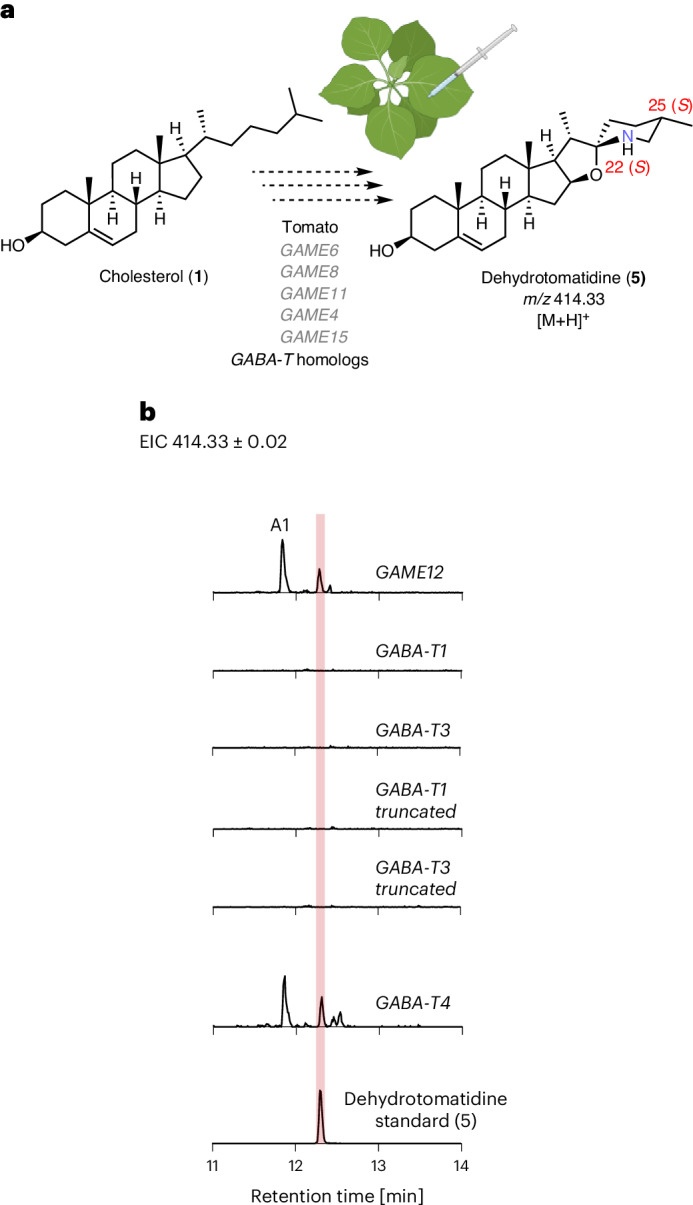
Steroidal glycoalkaloids (SGAs) are specialized metabolites produced by hundreds of Solanum species including food crops, such as tomato, potato and eggplant. Unlike true alkaloids, nitrogen is introduced at a late stage of SGA biosynthesis through an unknown transamination reaction. Here, we reveal the mechanism by which GLYCOALKALOID METABOLISM12 (GAME12) directs the biosynthesis of nitrogen-containing steroidal alkaloid aglycone in Solanum. We report that GAME12, a neofunctionalized γ-aminobutyric acid (GABA) transaminase, undergoes changes in both active site specificity and subcellular localization to switch from its renown and generic activity in core metabolism to function in a specialized metabolic pathway. Moreover, overexpression of GAME12 alone in engineered S. nigrum leaves is sufficient for de novo production of nitrogen-containing SGAs. Our results highlight how hijacking a core metabolism GABA shunt enzyme is crucial in numerous Solanum species for incorporating a nitrogen to a steroidal-specialized metabolite backbone and form defensive alkaloids.


Conflict of interest statement

Competing interests: The authors declare no competing interests.

Figures

Fig. 1
Fig. 1. Predicted biosynthetic pathway of SGAs in Solanum species.
a, The predicted biosynthetic pathway of SGAs in potato (S.tuberosum), tomato (S.lycopersicum) and eggplant (S.melongena). Solid arrows represent known biosynthetic steps and dashed arrows show uncharacterized steps. Unconfirmed intermediates in the SGA pathway are placed in brackets. Colored arrows represent the branches of SGA biosynthesis specific to different Solanum species. The relevant carbon positions modified by the biosynthetic enzymes are numbered in red. b, The primary metabolism GABA-T catalyzes pyruvate (10) transamination, leading to the formation of alanine (11) and succinic semialdehyde (13), using GABA (12) as a cosubstrate. Gal, galactose; Glc, glucose; Rha, rhamnose; Xyl, xylose; DPS, dioxygenase for potato solanidane synthesis; 5αR2, steroid 5α-reductase 2.
Fig. 2
Fig. 2. Activity of the tomato GABA-T homologs transiently expressed in N.benthamiana.
a, Schematic representation of the pathway reconstitution approach used to assay the GABA-T homologs in N.benthamiana. b, Extracted ion chromatograms (EICs) showing the products of the pathway reconstitution-based assays and the corresponding dehydrotomatidine (5) standard. The MS2 spectra of the products and the dehydrotomatidine standard (5) are presented in Supplementary Fig. 3. The scale is uniform across all chromatograms (the y axis signifies signal intensity, where the maximum is 1.6 × 104), except for the dehydrotomatidine (5) standard chromatogram where the y axis maximum is 1.6 × 105).
Fig. 3
Fig. 3. Establishment of in vitro assays of tomato GAME4 and GAME12 leading to the production of steroidal alkaloid aglycones solasodine (6) and soladulcidine (17).
a, The catalytic steps occurring in the in vitro assays. Both unsaturated (14) and saturated (16) (C-5,6 bond marked with a dashed red line) steroidal saponins can serve as GAME4 and GAME12 substrates after hydrolysis. b, EICs of the in vitro assays products using protodioscin (14) as a substrate and the corresponding solasodine (6) standard. c, EICs of the in vitro assays products using uttroside B (16) as a substrate and the corresponding soladulcidine (17) standard. The scale is uniform across all of the chromatograms (the y axis signifies signal intensity, where the maximum is 1.6 × 104). The MS2 spectra of the products and standards are presented in Supplementary Fig. 7.
Fig. 4
Fig. 4. The activity of the tomato GABA-T homologs in in vitro coupled assays and mutagenesis of GABA-T3 for the gain of SGA-forming activity.
a, EICs of the Rapidase–GAME4-coupled in vitro assays using protodioscin (14) as a substrate and the corresponding solasodine (6) standard. For the characterization of the additional product A2, a putative 26-aminofurostanol (Supplementary Fig. 7), the concentration of proteins in the assay was normalized to 1 μM of purified protein for all GABA-T homologs. The scale is uniform across all chromatograms. b, Visualization of the AlphaFold-generated model of the GAME12 homodimer with 26-furostanol aldehyde (3) docked into the active site and the PMP cofactor modeled in using a previously solved crystal structure of a GABA-T (PDB 4ATQ), as described in the Methods,. The model represents the beginning of the second transamination half-reaction, where the keto acid substrate (26-furostanol aldehyde (3)) acts as an amine acceptor. The GAME12 residues corresponding to the codons identified to be under strong diversifying selection are labeled in orange boxes. c, Sequence alignment of GAME12, GABA-T3 and GABA-T3 mut2 and mut3. Residue numbering is consistent with the diversifying selection analysis codon numbering. The residues corresponding to the codons identified to be under strong diversifying selection are underlined in orange. The GABA-T3 mut2 and mut3 residues highlighted in color were substituted with the corresponding residues from GAME12. d, EICs of the Rapidase–GAME4-coupled in vitro assays of the active GABA-T3 mutants using protodioscin (14) as a substrate. The concentration of proteins in the assay was normalized to 1 μM of total protein for all GABA-T homologs. The scale is uniform across all chromatograms of a and d (the y axis signifies signal intensity, where the maximum is 1.6 × 104), except for the ×10 magnification on the chromatogram in d marked with a gray box where the y axis maximum of 1.6 × 103.
Fig. 5
Fig. 5. Introducing nitrogen into a steroidal backbone in stably transformed S.nigrum plants.
a, Proposed biosynthesis of the most abundant furostane-type steroidal saponin, uttroside B (16) in S.nigrum leaves and the putatively assigned structures of soladulcidine (17)-type SGAs extracted from wild-type S.nigrum green berries. The early biosynthetic steps up to the formation of furostanol (2) are hypothesized to be common between the steroidal saponin and SGA pathways in different Solanum species. MS2-based putative structural assignment of the de novo produced SGAs (peaks D, E and F) can be found in Supplementary Table 5. b, EICs from transgenic and wild-type S.nigrum lines. S.nigrum leaves stably transformed with GAME12 produce SGAs, as shown by the presence of the characteristic m/z 416.35, corresponding to steroidal alkaloid aglycone soladulcidine (17) observed in UHPLC–MS analysis. The scale is uniform across all chromatograms (the y axis signifies signal intensity, where the maximum is 1 × 106) c, The MS2 spectrum of product E, displaying the ion of m/z 416.35, characteristic to SGA scaffolds containing a soladulcidine (17)-type core. d, Comparison of the peak areas of product E, a de novo produced SGA, and the natively produced furostanol-type steroidal saponin uttroside B (16) in the wild-type S.nigrum plants, tomato GAME12-overexpressing S.nigrum transgenic lines (3, 6, 19 and 20) and S.nigrum lines (14 and 16) stably transformed with tomato GAME4 and GAME12. The bar graphs represent the mean ± s.d. for three biological replicates (n = 3). Asterisks signify a statistically significant difference in the area of the uttroside B (16) peak between the transgenic lines and wild-type S.nigrum plants according to an unpaired, two-tailed t-test (for line 14, *P = 0.0029; for line 16, *P = 0.038). Source data
Fig. 6
Fig. 6. Model of the evolution of the SGA-forming GAME12 enzyme from the canonical GABA-Ts of the Solanaceae.
a, Model of GABA-T evolution in the Solanaceae. GABA-T homologs (arrows) are colored according to the clades in Extended Data Fig. 1a, with the color of the squares matching the syntenic regions in Extended Data Fig. 1b. The square of the C.annuum VAMT is black, as it is located on an unrelated locus outside of the syntenic regions mentioned in Extended Data Fig. 1b. Selected reported whole-genome duplication events are labeled with a red star and loss of GABA-T4 is labeled with a gray star. b, Simplified depiction of the changes at the branch points of the evolution of GABA-T homologs in S.lycopersicum.
Extended Data Fig. 1
Extended Data Fig. 1. Phylogenomic analysis of GABA-Ts.
a. Maximum likelihood gene tree of GABA-T homologues. Thick branches show those selected for diversifying selection tests. Branches are coloured if significant selection was detected, with color showing ω1 rate class value in model with one rate class per branch. Grey circles show branches with >80% and >95% support as judged by 1000X SH-aLRT and UltraFast Boostrapping replicates, respectively. An extended phylogentic tree can be found in Supplementary Fig. 25. b. Microsynteny analysis of GABA-T homologues. Curved lines connecting genomes represent homologous genes, with colors added manually to track genes of interest. Gaps (10-70 Mb) in synteny represented with double slash. Pseudogenes (genes that have premature stop codons in exons, highly divergent sequences or missing expected coding regions. are marked with the Ψ symbol. Larger versions of the results of each syntenic block analysis in panel (b) can be found in Supplementary Fig. 26. Source data

Similar articles

Cited by

References

    1. Schreiber, K. in The Alkaloids: Chemistry and Physiology (ed. Manske, R. H. F.) (Academic Press, 1968).
    1. Roddick, J. G. in Saponins Used in Traditional and Modern Medicine (eds Waller, G. R. & Yamasaki, K.) (Springer, 1996).
    1. Lichman, B. R. The scaffold-forming steps of plant alkaloid biosynthesis. Nat. Prod. Rep.38, 103–129 (2021). - PubMed
    1. Itkin, M. et al. Biosynthesis of antinutritional alkaloids in solanaceous crops is mediated by clustered genes. Science341, 175–179 (2013). - PubMed
    1. Itkin, M. et al. GLYCOALKALOID METABOLISM1 is required for steroidal alkaloid glycosylation and prevention of phytotoxicity in tomato. Plant Cell23, 4507–4525 (2011). - PMC - PubMed

Publication types

MeSH terms

LinkOut - more resources