Turkish coffee has an antitumor effect on breast cancer cells in vitro and in vivo
- PMID: 39272080
- PMCID: PMC11396339
- DOI: 10.1186/s12986-024-00846-4
Turkish coffee has an antitumor effect on breast cancer cells in vitro and in vivo
Abstract
Background: Breast cancer is the most diagnosed cancer in women. Its pathogenesis includes several pathways in cancer proliferation, apoptosis, and metastasis. Some clinical data have indicated the association between coffee consumption and decreased cancer risk. However, little data is available on the effect of coffee on breast cancer cells in vitro and in vivo.
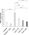
Methods: In our study, we assessed the effect of Turkish coffee and Fridamycin-H on different pathways in breast cancer, including apoptosis, proliferation, and oxidative stress. A human breast cancer cell line (MCF-7) was treated for 48 h with either coffee extract (5% or 10 v/v) or Fridamycin-H (10 ng/ml). Ehrlich solid tumors were induced in mice for in vivo modeling of breast cancer. Mice with Ehrlich solid tumors were treated orally with coffee extract in drinking water at a final concentration (v/v) of either 3%, 5%, or 10% daily for 21 days. Protein expression levels of Caspase-8 were determined in both in vitro and in vivo models using ELISA assay. Moreover, P-glycoprotein and peroxisome proliferator-activated receptor gamma (PPAR-γ) protein expression levels were analyzed in the in vitro model. β-catenin protein expression was analyzed in tumor sections using immunohistochemical analysis. In addition, malondialdehyde (MDA) serum levels were analyzed using colorimetry.
Results: Both coffee extract and Fridamycin-H significantly increased Caspase-8, P-glycoprotein, and PPAR-γ protein levels in MCF-7 cells. Consistently, all doses of in vivo coffee treatment induced a significant increase in Caspase-8 and necrotic zones and a significant decrease in β- catenin, MDA, tumor volume, tumor weight, and viable tumor cell density.
Conclusion: These findings suggest that coffee extract and Fridamycin-H warrant further exploration as potential therapies for breast cancer.
Keywords: Breast cancer; Caspase-8; Coffee extract; Fridamycin-H; PPAR-γ; Β-catenin.
© 2024. The Author(s).
Conflict of interest statement
The authors declare no competing interests.
Figures


Similar articles
-
Antitumor Effects of Freeze-Dried Robusta Coffee (Coffea canephora) Extracts on Breast Cancer Cell Lines.Oxid Med Cell Longev. 2021 May 18;2021:5572630. doi: 10.1155/2021/5572630. eCollection 2021. Oxid Med Cell Longev. 2021. PMID: 34113419 Free PMC article.
-
Caspase-dependent and caspase-independent induction of apoptosis in breast cancer by fucoidan via the PI3K/AKT/GSK3β pathway in vivo and in vitro.Biomed Pharmacother. 2017 Oct;94:898-908. doi: 10.1016/j.biopha.2017.08.013. Epub 2017 Aug 12. Biomed Pharmacother. 2017. PMID: 28810530
-
Coffee component hydroxyl hydroquinone (HHQ) as a putative ligand for PPAR gamma and implications in breast cancer.BMC Genomics. 2013;14 Suppl 5(Suppl 5):S6. doi: 10.1186/1471-2164-14-S5-S6. Epub 2013 Oct 16. BMC Genomics. 2013. Retraction in: BMC Genomics. 2022 Feb 14;23(1):127. doi: 10.1186/s12864-022-08371-5. PMID: 24564733 Free PMC article. Retracted.
-
Dendropanax morbiferus H. Lév. Leaf Extract Inhibits the Proliferation of MDA-MB-231 Breast Cancer Cells and Induces Apoptosis via the MAPK Pathway In Vitro and In Vivo.Anticancer Res. 2023 Jul;43(7):3047-3056. doi: 10.21873/anticanres.16476. Anticancer Res. 2023. PMID: 37351981
-
Salvianolic Acid B Slows the Progression of Breast Cancer Cell Growth via Enhancement of Apoptosis and Reduction of Oxidative Stress, Inflammation, and Angiogenesis.Int J Mol Sci. 2019 Nov 12;20(22):5653. doi: 10.3390/ijms20225653. Int J Mol Sci. 2019. PMID: 31726654 Free PMC article.
References
LinkOut - more resources
Full Text Sources

