. 2024 Sep 13;16(1):112.
doi: 10.1186/s13073-024-01378-5.

Chromatin conformation capture in the clinic: 4C-seq/HiC distinguishes pathogenic from neutral duplications at the GPR101 locus

Affiliations

Chromatin conformation capture in the clinic: 4C-seq/HiC distinguishes pathogenic from neutral duplications at the GPR101 locus

Adrian F Daly et al. Genome Med. .

Abstract

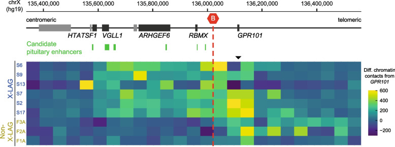
Background: X-linked acrogigantism (X-LAG; MIM: 300942) is a severe form of pituitary gigantism caused by chromosome Xq26.3 duplications involving GPR101. X-LAG-associated duplications disrupt the integrity of the topologically associating domain (TAD) containing GPR101 and lead to the formation of a neo-TAD that drives pituitary GPR101 misexpression and gigantism. As X-LAG is fully penetrant and heritable, duplications involving GPR101 identified on prenatal screening studies, like amniocentesis, can pose an interpretation challenge for medical geneticists and raise important concerns for patients and families. Therefore, providing robust information on the functional genomic impact of such duplications has important research and clinical value with respect to gene regulation and triplosensitivity traits.

Methods: We employed 4C/HiC-seq as a clinical tool to determine the functional impact of incidentally discovered GPR101 duplications on TAD integrity in three families. After defining duplications and breakpoints around GPR101 by clinical-grade and high-density aCGH, we constructed 4C/HiC chromatin contact maps for our study population and compared them with normal and active (X-LAG) controls.

Results: We showed that duplications involving GPR101 that preserved the centromeric invariant TAD boundary did not generate a pathogenic neo-TAD and that ectopic enhancers were not adopted. This allowed us to discount presumptive/suspected X-LAG diagnoses and GPR101 misexpression, obviating the need for intensive clinical follow-up.

Conclusions: This study highlights the importance of TAD boundaries and chromatin interactions in determining the functional impact of copy number variants and provides proof-of-concept for using 4C/HiC-seq as a clinical tool to acquire crucial information for genetic counseling and to support clinical decision-making in cases of suspected TADopathies.

Keywords: 4C; Chromosome microarray; Enhancer; GPR101; HiC; Neo-TAD; Pituitary tumor; Prenatal diagnosis; Topologically associating domains; X-linked acrogigantism.


Conflict of interest statement

AFD, FRF, AB, CAS, and GT hold a patent on GPR101 and its function (US Patent No. 10,350,273, Treatment of Hormonal Disorders of Growth). JRL has stock ownership in 23andMe and is a paid consultant for Genome International. CAS is co-founder and Director of ASTREA, a precision medicine company. The authors declare no other competing interests.

Figures

Fig. 1
Fig. 1
Impact of inter- and intra-TAD duplications on chromatin organization at the X-LAG locus. A Schematic representation of the extended X-LAG locus (hg19, chrX:135,336,766–136,561,684), delineating the genomic position of an invariant TAD border (red hexagon). Below, the position of partially overlapping tandem duplications involving GPR101 from subjects with X-LAG (highlighted by blue boxes) [16] that traverse the TAD border (inter-TAD duplications), alongside duplications from subjects of the current study (yellow boxes) that remain within TAD boundaries (intra-TAD duplications). B and C HiC at the X-LAG locus, showing normalized contact matrices at 10-kb resolution from the X-LAG subject S7 and non-X-LAG subject F2A in a side-by-side comparison to controls. Normal TAD configuration at the locus is highlighted by red arrows. Additional chromatin interactions, induced by inter- and intra-TAD duplications, are denoted by black arrows. HiC difference maps relative to controls (D and E) depict the increase in chromatin interactions (black arrows) in subject S7 and F2A. Below, corresponding 4C-seq profiles originating from the GPR101 viewpoint (black triangle) are displayed, alongside the genomic position of the duplication and the subtraction profiles relative to control samples. Inter-TAD duplications in X-LAG (B and D) result in increased chromatin interaction of GPR101 with regions centromeric of the TAD border (neo-TAD formation). Intra-TAD duplications, confined to GPR101 and excluding the invariant TAD border (C and E), exhibit increased telomeric chromatin interactions
Fig. 2
Fig. 2
Modulation of GPR101 chromatin interactions by inter- and intra-TAD duplications. Schematic representation of the extended X-LAG locus (hg19, chrX:135,336,766–136,561,684), delineating the genomic positions of putative pituitary enhancers (green boxes) and an invariant TAD border (red hexagon), which separates GPR101 from pituitary activity in normal conditions. Below, heatmap showing differential chromatin contacts at a 50-kb genomic bin size in X-LAG and non-X-LAG subject samples compared to controls, as inferred from 4C-seq experiments with a viewpoint located at the GPR101 promoter. The genomic bin containing the viewpoint is indicated by a black arrowhead. All X-LAG subjects exhibit similar patterns of ectopic chromatin interactions from GPR101 with the centromeric region containing putative pituitary enhancers. This pattern is absent in non-X-LAG subjects. The 4C-seq data from X-LAG subjects S6, S9, S13, S7, S2, and S17 and their respective controls (described in Franke et al. [16]) were retrieved from the GEO database under accession code GSE193114 [29]
Fig. 3
Fig. 3
Disease mechanism induced by inter- and intra-TAD duplications at the X-LAG locus. Schematic illustrating the configuration of TAD boundaries at the X-LAG locus with a linear genomic view (left) and a schematic representation of TAD configuration (right). A Under normal conditions, GPR101 is separated by a TAD boundary (red hexagon) from putative pituitary enhancers (green ovals). B Inter-TAD duplications associated with X-LAG, spanning the TAD boundary, lead to the formation of a neo-TAD (blue) that involves ectopic chromatin interactions between GPR101 and pituitary enhancers, consequently causing GPR101 misexpression and gigantism. C Intra-TAD duplications of GPR101 that “preserve” the TAD boundary do not generate a neo-TAD. As a result, the additional copy of GPR101 remains segregated from pituitary enhancers. Note that the size and position of duplications are indicated by overlap in the schematic representation

References

    1. Ibrahim DM, Mundlos S. Three-dimensional chromatin in disease: what holds us together and what drives us apart? Curr Opin Cell Biol. 2020;64:1–9. 10.1016/j.ceb.2020.01.003. 10.1016/j.ceb.2020.01.003 - DOI - PubMed
    1. da Costa-Nunes JA, Noordermeer D. TADs: dynamic structures to create stable regulatory functions. Curr Opin Struct Biol. 2023;81:102622. 10.1016/j.sbi.2023.102622. 10.1016/j.sbi.2023.102622 - DOI - PubMed
    1. Dixon JR, Selvaraj S, Yue F, Kim A, Li Y, Shen Y, et al. Topological domains in mammalian genomes identified by analysis of chromatin interactions. Nature. 2012;485:376–80. 10.1038/nature11082. 10.1038/nature11082 - DOI - PMC - PubMed
    1. Phillips-Cremins JE, Sauria MEG, Sanyal A, Gerasimova TI, Lajoie BR, Bell JSK, et al. Architectural protein subclasses shape 3D organization of genomes during lineage commitment. Cell. 2013;153:1281–95. 10.1016/J.CELL.2013.04.053. 10.1016/J.CELL.2013.04.053 - DOI - PMC - PubMed
    1. Lupiáñez DG, Spielmann M, Mundlos S. Breaking TADs: how alterations of chromatin domains result in disease. Trends Genet. 2016;32:225–37. 10.1016/J.TIG.2016.01.003. 10.1016/J.TIG.2016.01.003 - DOI - PubMed