. 2024 Sep 16;405(11-12):711-726.
doi: 10.1515/hsz-2024-0088. Print 2024 Dec 17.

The BCL11A transcription factor stimulates the enzymatic activities of the OGG1 DNA glycosylase

Affiliations

The BCL11A transcription factor stimulates the enzymatic activities of the OGG1 DNA glycosylase

Tetiana Petrachkova et al. Biol Chem. .

Abstract

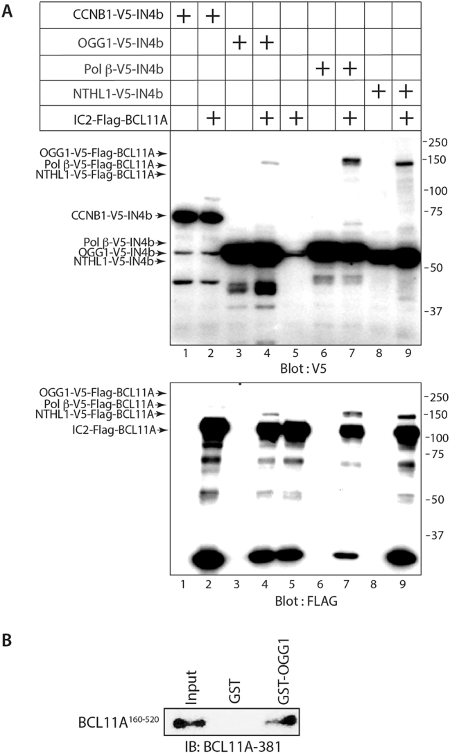
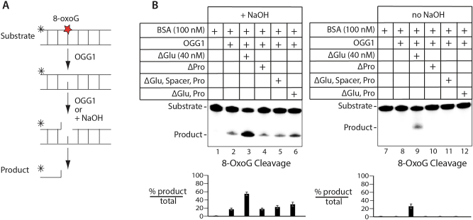
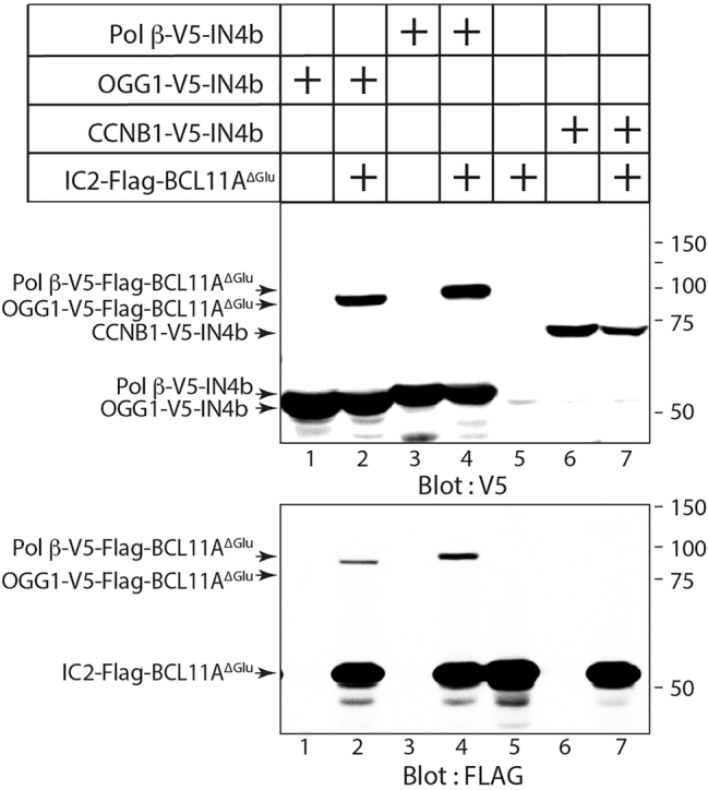
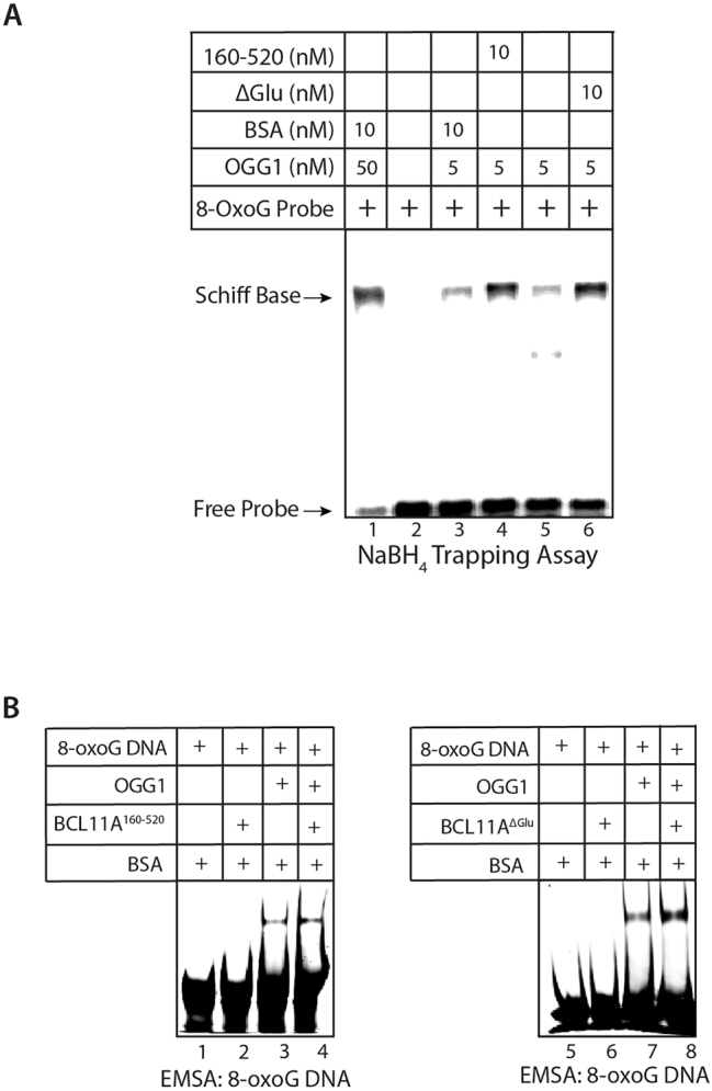
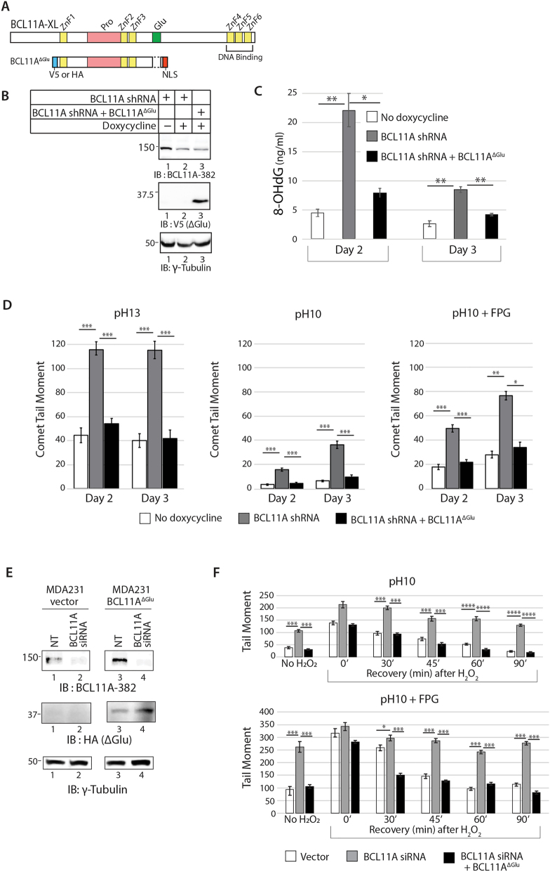
The BCL11A transcription factor has previously been shown to interact with and stimulate the enzymatic activities of the NTHL1 DNA glycosylase and Pol β polymerase. Here we show that BCL11A and a smaller peptide encompassing amino acids 160 to 520 can interact with the 8-oxoguanine DNA glycosylase, OGG1, increase the binding of OGG1 to DNA that contains an 8-oxoguanine base and stimulate the glycosylase activity of OGG1. Following BCL11A knockdown, we observed an increase in oxidized purines in the genome using comet assays, while immunoassays reveal an increase in 8-oxoG bases. Structure-function analysis indicates that the stimulation of OGG1 by BCL11A requires the zinc fingers 1, 2 and 3 as well as the proline-rich region between the first and second zing finger, but a glutamate-rich region downstream of zinc finger 3 is dispensable. Ectopic expression of a small peptide that contains the three zinc fingers can rescue the increase in 8-oxoguanine caused by BCL11A knockdown. These findings, together with previous results showing that BCL11A stimulates the enzymatic activities of NTHL1 and the Pol β polymerase, suggest that high expression of BCL11A is important to protect cancer cells against oxidative DNA damage.

Keywords: 8-oxoguanine glycosylase (OGG1); BCL11A; base excision repair; breast cancer; reactive oxygen species (ROS); transcription factor.


Conflict of interest statement

Competing interests: The authors state no conflict of interest.

Figures

Figure 1:
Figure 1:
BCL11A stimulates OGG1 in vitro and regulates oxidized purines in cells. (A) Map of the full-length BCL11A protein and the BCL11A160–520 protein that were tested in the 8-oxoG cleavage assay. (B) MDA-MB-231 cells were stably infected with the pCF806 lentivirus vector expressing BCL11A shRNA under the control of a doxycycline-inducible promoter. Doxycycline was added to the medium or not and after 3 days immunoblotting was performed with the BCL11A-382 antibody and ɣ-tubulin. Single cell gel electrophoresis was performed at pH > 13, pH 10 and pH 10 after treatment with the FPG DNA glycosylase. Comet tail moments were scored for at least 100 cells per condition. Results are a representative of one of three different experiments. Error bars represent standard error. ***p < 0.001; **p < 0.01; *p < 0.05; Student’s t-test. (C) Diagrammatic representation of the 8-oxoG cleavage assay using a fluorophore reporter probe. The assay was performed using 5 pmol of the 8-oxoG probe, 10 nM of OGG1, 10 nM of the BCL11A-XL, or 40 nM of the BCL11A160–520, in the presence of 100 nM of BSA; reaction volume 20 μL. Error bars represent standard deviation. (D) MDA-MB-231 cells were infected with lentiviral vectors as indicated: an empty vector or a vector expressing HA-BCL11A160–520. Cells were transfected with either non-targeting siRNA (NT siRNA), BCL11A siRNA1 or siRNA2. Immunoblotting was performed with the BCL11A-382 antibody and an HA antibody for the HA-BCL11A160–520 protein. Cells were treated with 50 µM H2O2 for 20 min or not (non-treated), followed by either a recovery period of 45 min or no recovery (0 min). Single cell gel electrophoresis was performed at pH 10 and pH 10 after treatment with the FPG DNA glycosylase. Comet tail moments were scored for at least 100 cells per condition. Results are a representative of one of three different experiments. Error bars represent standard error. ***p < 0.001; **p < 0.01; *p < 0.05; Student’s t-test.
Figure 2:
Figure 2:
BCL11A interacts with the OGG1 DNA glycosylase. (A) Split-Intein Assay: 293 cells were transfected with vectors expressing fusion proteins containing either the N-terminal or C-terminal portion of Intein, as indicated: IC2-Flag-BCL11A, OGG1-V5-IN4b, Pol β-V5-IN4B, NTHL1-V5-IN4B or CCNB1-V5-IN4B. Whole cell extracts were submitted to immunoblotting analysis with the V5 and FLAG antibodies. Recombinant proteins are observed in lanes 4, 7 and 9, but not in lane 2. (B) Pull-down assays were performed using the BCL11A160–520 protein fragment and either GST or GST-OGG1, followed by immunoblotting with anti-BCL11A-381 antibody.
Figure 3:
Figure 3:
Structure-function analysis of BCL11A defines a region of BCL11A that stimulates the enzymatic activity of OGG1. (A) Diagrammatic representation of the BCL11A peptides tested in 8-oxoG assays. Coomassie blue stain of the BCL11A peptides purified from bacteria. (B) Diagrammatic representation of the 8-oxoG cleavage assay using a fluorophore reporter probe. (C) The assay was performed using 5 pmol of the 8-oxoG probe, 10 nM of OGG1 or 20 nM of the BCL11A peptides in the absence of OGG1; reaction volume 20 μL. (D–F) The assay was performed using 5 pmol of the 8-oxoG probe, 10 nM of OGG1 and 20 nM of the BCL11A peptides, in the presence of 100 nM of BSA; reaction volume 20 μL. Error bars represent standard deviation. (G) The histogram shows the fold change in 8-oxoG cleavage observed in the presence of various BCL11A peptides.
Figure 4:
Figure 4:
The ∆Glu BCL11A peptide stimulates the enzymatic activity of OGG1. (A) Diagrammatic representation of the 8-oxoG cleavage assay using a radioactively labeled probe. (B) 8-oxoG cleavage assays were performed using radioactively end-labeled double-stranded oligonucleotides containing an 8-oxoG base, and 100 nM BSA; reaction volume 20 μL. Where indicated, OGG1 (2.5 nM), and some BCL11A peptides (40 nM) were included in the reaction. Production of a cleaved product in samples that have not been treated with NaOH (lanes 7 to 12) requires the glycosylase and AP/lyase activities of OGG1.
Figure 5:
Figure 5:
BCL11A∆Glu interacts with the OGG1 DNA glycosylase. Split-Intein Assay: 293 cells were transfected with vectors expressing fusion proteins containing either the N-terminal or C-terminal portion of Intein, as indicated: IC2-Flag-BCL11A∆Glu, Pol β-V5-IN4B, OGG1-V5-IN4b or CCNB1-V5-IN4B. Whole cell extracts were submitted to immunoblotting analysis with the V5 and FLAG antibodies. Recombinant proteins are observed in lanes 2 and 4, but not in lane 7.
Figure 6:
Figure 6:
The 160–520 and ∆Glu BCL11A peptides stimulate the binding of OGG1 to DNA containing an 8-oxoG. (A) A Schiff base assay was performed using a radioactively labeled probe containing an 8-oxoG residue. The probes were incubated with OGG1 in the presence of BCL11A160–520, BCL11A∆Glu, or BSA (10 nM); reaction volume 30 μL following the incubation, 50 mM sodium borohydride was added. The trapped Schiff base intermediates were separated from the free probe by 10 % SDS-PAGE. (B) EMSA was performed with double-stranded oligonucleotides containing an 8-oxoG base, OGG1 (20 nM), either alone or with BCL11A160–520 (10 nM) or with BCL11A∆Glu (10 nM); reaction volume 20 μL. The proteins were incubated with the probe for 3 min.
Figure 7:
Figure 7:
Ectopic expression of the ∆Glu BCL11A160–480 Peptide rescues the increase in oxidative DNA damage caused by BCL11A knockdown. (A) Schematic representation of the BCL11A-XL and BCl11AΔGlu proteins. (B–D) MDA-MB-231 cells were stably infected with the pCF806 lentiviral vector expressing BCL11A shRNA or BCL11A shRNA and V5-BCL11A∆Glu under the control of a doxycycline-inducible promoter. (B) Doxycycline was added or not to the medium and two days later immunoblotting was performed with anti-BCL11A-382 antibody for the endogenous BCL11A, and anti-V5 antibody for the BCL11AΔGlu-V5 protein. (C) 8-OHdG levels were measured on day 2 and 3 after doxycycline induction. The values are the mean of three measurements and error bars represent standard deviation, Student’s t-test, ***p < 0.001; **p < 0.01; *p < 0.05. (D) On day 2 and 3 after doxycycline induction, single-cell gel electrophoresis was performed at pH > 13, pH 10 and pH 10 after treatment with the FPG DNA glycosylase. Comet tail moments were scored for at least 100 cells per condition. Error bars represent standard error. Student’s t-test, ***p < 0.001; **p < 0.01; *p < 0.05, ns, not significant. (E and F) MDA-MB-231 cells were stably infected with plenti6 lentiviral vectors constitutively expressing either nothing (Vector) or HA-BCL11A∆Glu. Cells were then transfected with either non-targeting siRNA (NT siRNA) or BCL11A siRNA. (E) Two days following transfection, immunoblotting was performed with the BCL11A-382 antibody and an HA antibody for the HA-BCL11A∆Glu protein. (F) Two days following transfection, cells were treated or not with 50 µM H2O2 and allowed to recover for the indicated periods of time. Single-cell gel electrophoresis was performed at pH 10 and pH 10 after treating cells with the FPG glycosylase. Comet tail moments were scored for at least 100 cells per condition. Error bars represent standard error. Student’s t-test, ***p < 0.001; **p < 0.01; *p < 0.05, ns, not significant.

Similar articles

References

    1. Amen A.M., Loughran R.M., Huang C.H., Lew R.J., Ravi A., Guan Y., Schatoff E.M., Dow L.E., Emerling B.M., Fellmann C. Endogenous spacing enables co-processing of microRNAs and efficient combinatorial RNAi. Cell Rep. Methods . 2022;2:100239. doi: 10.1016/j.crmeth.2022.100239. - DOI - PMC - PubMed
    1. Avram D., Fields A., Pretty On Top K., Nevrivy D.J., Ishmael J.E., Leid M. Isolation of a novel family of C(2)H(2) zinc finger proteins implicated in transcriptional repression mediated by chicken ovalbumin upstream promoter transcription factor (COUP-TF) orphan nuclear receptors. J. Biol. Chem. . 2000;275:10315–10322. doi: 10.1074/jbc.275.14.10315. - DOI - PMC - PubMed
    1. Basu T.N., Gutmann D.H., Fletcher J.A., Glover T.W., Collins F.S., Downward J. Aberrant regulation of ras proteins in malignant tumour cells from type 1 neurofibromatosis patients. Nature . 1992;356:713–715. doi: 10.1038/356713a0. - DOI - PubMed
    1. Cadieux C., Kedinger V., Yao L., Vadnais C., Drossos M., Paquet M., Nepveu A. Mouse mammary tumor virus p75 and p110 CUX1 transgenic mice develop mammary tumors of various histologic types. Cancer Res. . 2009;69:7188–7197. doi: 10.1158/0008-5472.can-08-4899. - DOI - PubMed
    1. Chakraborty A., Tapryal N., Islam A., Mitra S., Hazra T. Transcription coupled base excision repair in mammalian cells: so little is known and so much to uncover. DNA Repair . 2021;107:103204. doi: 10.1016/j.dnarep.2021.103204. - DOI - PubMed

Publication types

MeSH terms

LinkOut - more resources