. 2024 Aug 28;14(17):2500.
doi: 10.3390/ani14172500.

RNA-Seq Analysis of Glycolysis Regulation of Avian Leukosis Virus Subgroup J Replication

Affiliations

RNA-Seq Analysis of Glycolysis Regulation of Avian Leukosis Virus Subgroup J Replication

Ting Yang et al. Animals (Basel). .

Abstract

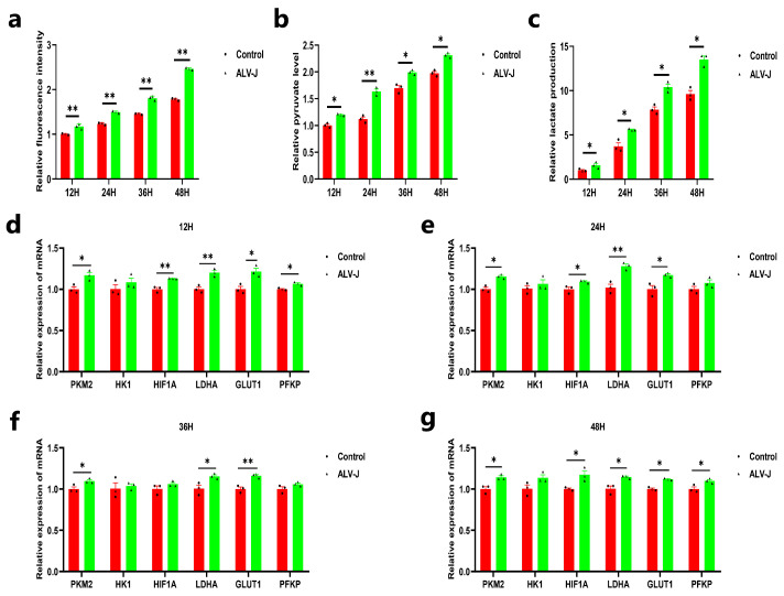
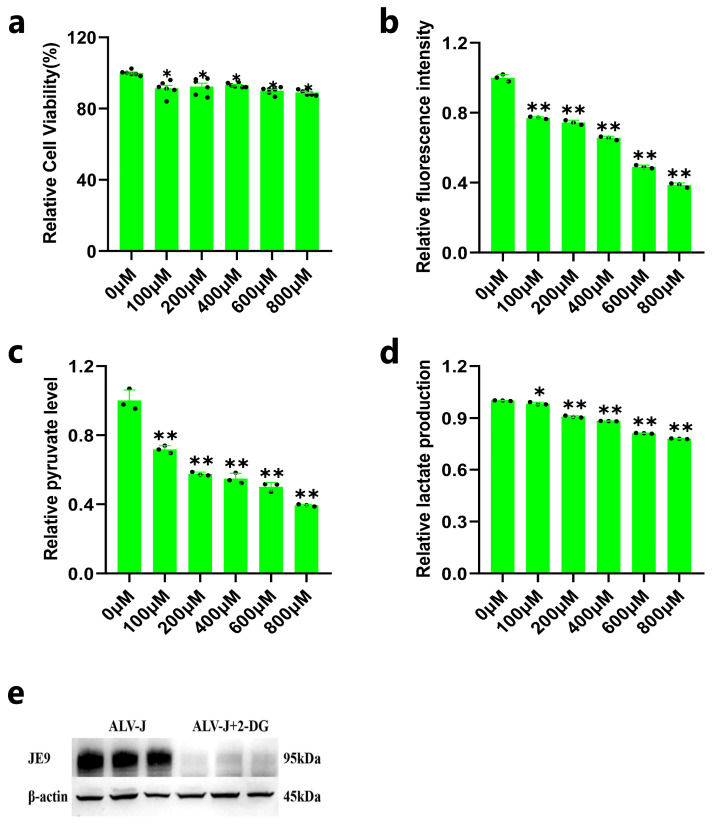
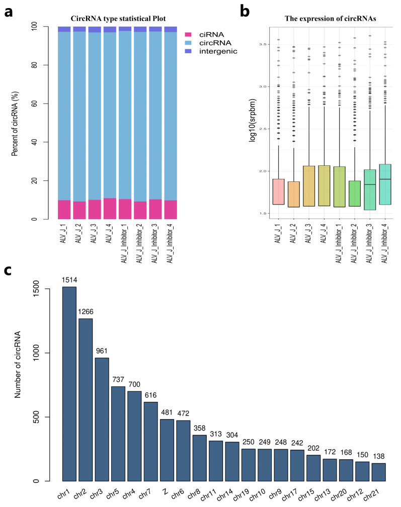
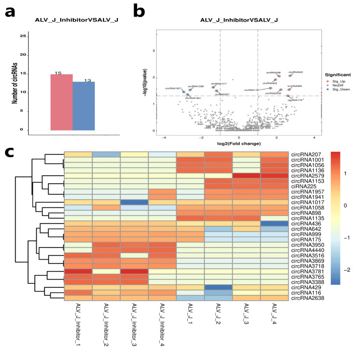
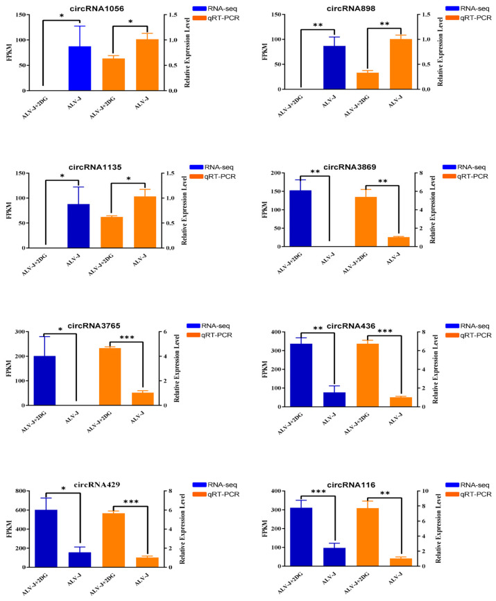
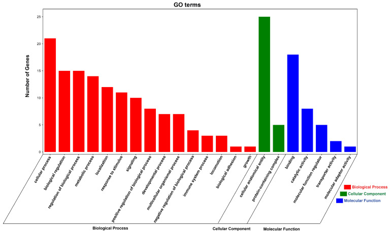
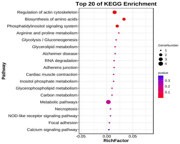
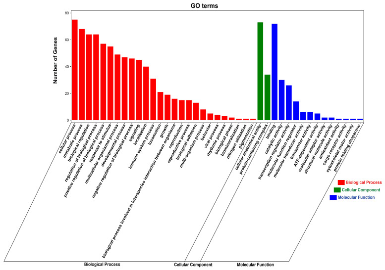
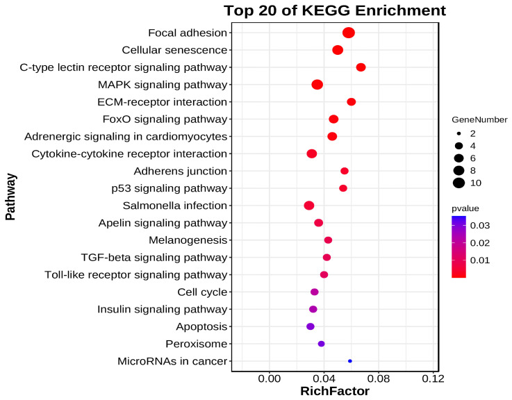
Avian Leukosis virus (ALV) is a widely spread virus that causes major economic losses to the global poultry industry. This study aims to investigate the effect of glycolysis on the replication of the ALV-J virus and identify the key circular RNAs that regulate the replication of the ALV-J virus. We found that glucose uptake, pyruvate content, and lactate content in DF1 cells were increased after ALV-J infection. Moreover, inhibiting the glycolysis of ALV-J-infected DF1 cells reduced the replication of the ALV-J virus. To further study the mechanism of glycolysis in the replication of the ALV-J virus, we performed RNA-seq on ALV-J-infected and ALV-J-infected cells treated with glycolysis inhibition. RNA-seq results show that a total of 10,375 circular RNAs (circRNAs) were identified, of which the main types were exonic circular RNAs, and 28 circRNAs were differentially expressed between ALV-J-infected and ALV-J-infected cells treated with glycolysis inhibition. Then, we performed functional enrichment analysis of differentially expressed circRNA source and target genes. Functional enrichment analysis indicated that some circRNAs might be involved in regulating the replication of the ALV-J virus by influencing some pathways like glycolysis/gluconeogenesis, the NOD-like receptor signaling pathway, MAPK signaling pathway, p53 signaling pathway, Toll-like receptor signaling pathway, Insulin signaling pathway, and Apoptosis. This study revealed the effect of glycolysis on the replication of the ALV-J virus in DF1 cells and its possible regulatory mechanism, which provided a basis for understanding the factors influencing the replication of the ALV-J virus and reducing the rate of infection of the ALV-J virus in poultry.

Keywords: ALV-J; CircRNA; glycolysis; virus replication.


Conflict of interest statement

The authors declare no conflicts of interest.

Figures

Figure 1
Figure 1
ALV-J elevates glucose uptake and glycolysis in DF1 cells. (a) The glucose uptake ability after DF1 cells were infected with the ALV-J virus. (b) The contents of pyruvate after DF1 cells were infected with the ALV-J virus. (c) The contents of lactate after DF1 cells were infected with the ALV-J virus. (dg) The mRNA expression level of glycolysis-related genes (PKM2, HK1, HIF1A, LDHA, GLUT1, PFKP) after DF1 cells were infected with the ALV-J virus. p < 0.05 (*) or p < 0.01 (**) represent statistically significant, and all data are shown as means ± SD (standard deviation).
Figure 2
Figure 2
Inhibition of DF1 cell glycolysis reduces ALV-J replication. (a) The cell viability after exposing DF1 cells to different concentrations of 2-DG. (b) The glucose uptake ability after exposing DF1 cells to different concentrations of 2-DG. (c) The contents of pyruvate after exposing DF1 cells to different concentrations of 2-DG. (d) The contents of lactate after exposing DF1 cells to different concentrations of 2-DG. (e) The protein expression of JE9 after ALV-J-infected DF1 cells was treated with a 100 µM concentration of 2-DG. p < 0.05 (*) or p < 0.01 (**) represent statistically significant, and all data are shown as means ± SD (standard deviation).
Figure 3
Figure 3
Overview of RNA sequencing data and identification of circular RNA. (a) Percent of identified circRNAs in various types. (b) The circRNA expression distribution for each sample. (c) The chromosomal location of the circRNAs.
Figure 4
Figure 4
Identification of DE circular RNAs. (a) Statistics on the number of DE Circular RNAs in the ALV-J inhibitor and the ALV-J group DF1 cells. (b) Volcano map of DE Circular RNAs expressed in the ALV-J inhibitor and the ALV-J group DF1 cells. The red dots represent upregulated circular RNAs; the blue dots represent downregulated circular RNAs and the grey dots represent no difference. (c) Heat map of the DE Circular RNAs in the ALV-J inhibitor and the ALV-J group DF1 cells.
Figure 5
Figure 5
qRT-PCR validation of differentially expressed circular RNAs. Blue represents RNA-seq results and orange represents qRT-PCR results. p < 0.05 (*), p < 0.01 (**), or p < 0.001 (***) represent statistically significant, and all data are shown as means ± SD (standard deviation).
Figure 6
Figure 6
GO enrichment analysis of DE circRNA source genes. The red column represents the Biological Process, the green column represents the Cellular Component and the blue column represents the Molecular Function.
Figure 7
Figure 7
KEGG enrichment analysis of DE circRNA source genes. Top 20 pathway bubble diagram. Each bubble in Figure 7 represents a KEGG path, and the path name is shown in the legend on the left.
Figure 8
Figure 8
GO enrichment analysis of DE circRNA target genes. The red column represents the Biological Process, the green column represents the Cellular Component, and the blue column represents the Molecular Function.
Figure 9
Figure 9
KEGG Enrichment Analysis of DE circRNA Target Genes. Top 20 pathway bubble diagram. Each bubble in Figure 7 represents a KEGG path, and the path name is shown in the legend on the left.

References

    1. Feng M., Zhang X. Immunity to Avian Leukosis Virus: Where Are We Now and What Should We Do? Front. Immunol. 2016;7:624. doi: 10.3389/fimmu.2016.00624. - DOI - PMC - PubMed
    1. Dong X., Zhao P., Xu B., Fan J., Meng F., Sun P., Ju S., Li Y., Chang S., Shi W. Avian leukosis virus in indigenous chicken breeds, China. Emerg. Microbes Infect. 2015;4:1–2. doi: 10.1038/emi.2015.76. - DOI - PMC - PubMed
    1. Li X., Lin W., Chang S., Zhao P., Zhang X., Liu Y., Chen W., Li B., Shu D., Zhang H. Isolation, identification and evolution analysis of a novel subgroup of avian leukosis virus isolated from a local Chinese yellow broiler in South China. Arch. Virol. 2016;161:2717–2725. doi: 10.1007/s00705-016-2965-x. - DOI - PubMed
    1. Su Q., Li Y., Cui Z., Chang S., Zhao P. The emerging novel avian leukosis virus with mutations in the gene shows competitive replication advantages both in vivo and in vitro. Emerg. Microbes Infect. 2018;7:1–11. doi: 10.1038/s41426-018-0111-4. - DOI - PMC - PubMed
    1. Rubin H. The early history of tumor virology: Rous, RIF, and RAV. Proc. Natl. Acad. Sci. USA. 2011;108:14389–14396. doi: 10.1073/pnas.1108655108. - DOI - PMC - PubMed

LinkOut - more resources