. 2024 Aug 29;14(17):2519.
doi: 10.3390/ani14172519.

Pathologic Changes in and Immunophenotyping of Polymyositis in the Dutch Kooiker Dog

Affiliations

Pathologic Changes in and Immunophenotyping of Polymyositis in the Dutch Kooiker Dog

Vanessa Alf et al. Animals (Basel). .

Abstract

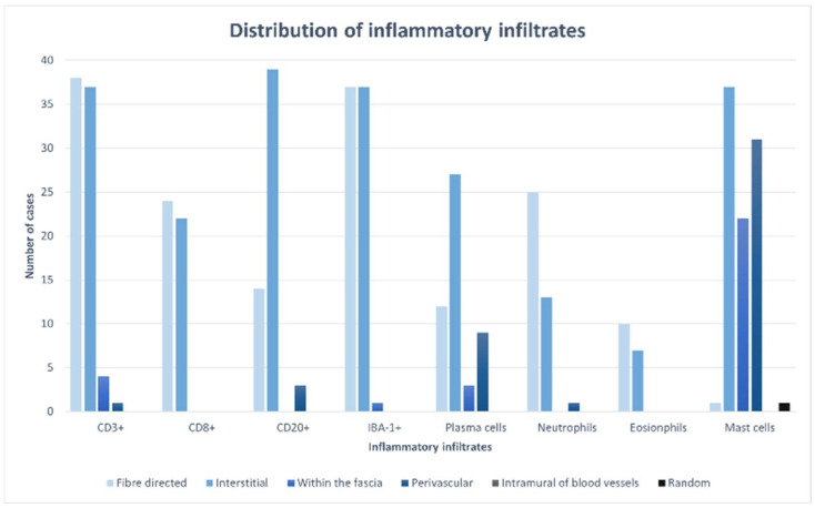
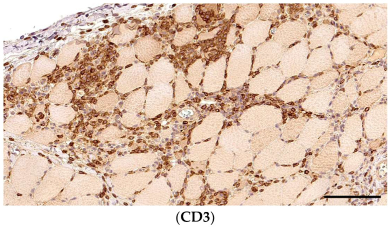
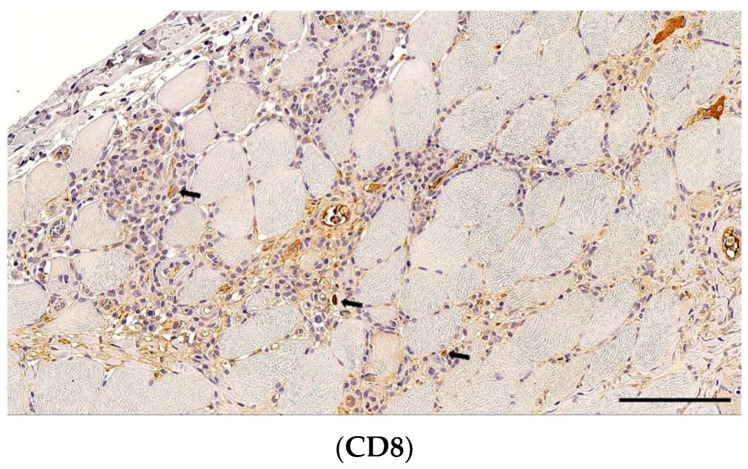
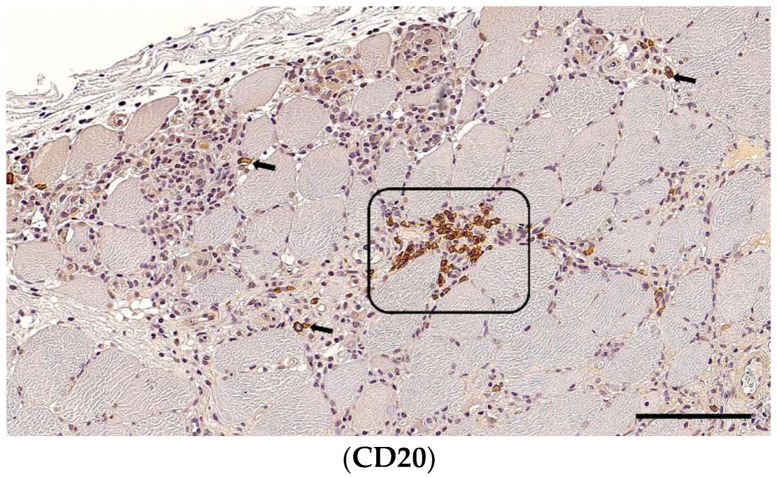
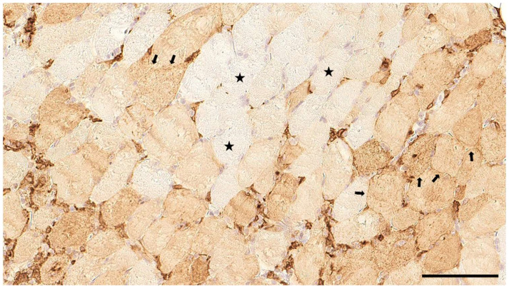
Earlier, we described a breed-specific inflammatory myopathy in Dutch Kooiker dogs (Het Nederlandse Kooikerhondje), one of the nine Dutch breeds. The disease commonly manifests itself with clinical signs of difficulty walking, muscle weakness, exercise intolerance, and/or dysphagia. In nearly all dogs' creatine kinase (CK) activity was elevated. Histopathology reveals the infiltration of inflammatory cells within the skeletal muscles. The objective of this study was to further investigate and characterize the histopathological changes in muscle tissue and immunophenotype the inflammatory infiltrates. FFPE fixed-muscle biopsies from 39 purebred Kooiker dogs were included and evaluated histopathologically according to a tailored classification scheme for skeletal muscle inflammation. As in other breed-related inflammatory myopathies, multifocal, mixed, and predominantly mononuclear cell infiltration was present, with an initial invasion of viable muscle fibres and the surrounding stroma leading to inflammation, necrosis, and tissue damage. Immunophenotyping primarily revealed lymphohistiocytic infiltrates, with CD3+ T-cells being the predominant inflammatory cell type, accompanied by CD8+ cytotoxic T-cells. The concurrent expression of MHC-II class molecules on myofibres suggests their involvement in initiating and maintaining inflammation. Additionally, CD20+ B-cells were identified, though in lower numbers compared to T-cells, and IBA-1-positive macrophages were frequently seen. These findings suggest a breed-specific subtype of polymyositis in Kooiker dogs, akin to other breeds. This study sheds light on the immune response activation, combining adaptive and innate mechanisms, contributing to our understanding of polymyositis in this breed.

Keywords: IBA-1; breed; hereditary disease; immune-mediated disease; inflammatory myopathy.


Conflict of interest statement

The authors declare no conflicts of interest.

Figures

Figure 1
Figure 1
Skeletal muscle of a Kooiker dog featuring a diffuse interstitial and myofibre-directed mononuclear cell infiltration consistent with myositis. Intact myocytes are infiltrated (coring out) by inflammatory cells (arrows). In more severely affected areas, varying stages of the myonecrosis of multiple fibres (frame) with the disruption of the sarcoplasm, invasion, and, later, replacement by macrophages being frequently seen. H&E, a section of skeletal muscle, marked severity, obj. 20×, scale bar = 100 µm.
Figure 2
Figure 2
Type and distribution of inflammatory infiltrates.
Figure 3
Figure 3
Immunohistochemistry for T-cells with fibre-directed and interstital distributions in the same section of skeletal muscle of Kooiker dogs. In CD3+ T-cells representing the predominant cell type in the Kooiker myositis, they surround and invade non-necrotic myofibres, characteristic of PM. A section of skeletal muscle, obj. 20×, scale bar = 100 µm.
Figure 4
Figure 4
Cytotoxic CD8+ cells scatter alongside the endomysium and in necrotic areas (arrows), albeit in lower number than CD3+ T-cells. A section of skeletal muscle, obj. 20×, scale bar = 100 µm.
Figure 5
Figure 5
Immunohistochemistry for B-cells and macrophages in the same skeletal muscle of a Kooiker dog. In contrast to previous reports, multifocal B-cell clusters (frame) and scattered B-cells (arrows) were frequently seen. CD20+ B-cells displayed interstitial infiltration and were concentrated in infiltrated and necrotic areas. A section of skeletal muscle, obj. 20×, scale bar = 100 µm.
Figure 6
Figure 6
Numerous IBA-1-expressing macrophages encompass non-necrotic myofibres and invade necrotic areas within damaged muscle tissue. A section of skeletal muscle, obj. 20×, scale bar = 100 µm.
Figure 7
Figure 7
MHC-II immunostaining in the skeletal muscle of a Kooiker dog. Besides APCs, multiple myofibres display a diffuse sarcolemmal MHC-II expression (arrows), whereas other myofibres are unremarkable (asterisks). A section of skeletal muscle, obj. 20×, scale bar = 100 µm.

Similar articles

Cited by

References

    1. Nielen A.L.J., van der Beek S., Ubbink G.J., Knol B.W. Epidemiology: Population parameters to compare dog breeds: Differences between five dutch purebred populations. Vet. Q. 2001;23:43–49. doi: 10.1080/01652176.2001.9695075. - DOI - PubMed
    1. Mandigers P.J., Van Nes J.J., Knol B.W., Ubbink G.J., Gruys E. Hereditary necrotising myelopathy in Kooiker dogs. Res. Vet. Sci. 1993;54:118–123. doi: 10.1016/0034-5288(93)90020-G. - DOI - PubMed
    1. Mandigers P.J., Ubbink G.J., Vanden Broek J., Bouw J. Relationship between litter size and other reproductive traits in the Dutch Kooiker dog. Vet. Q. 1994;16:229–232. doi: 10.1080/01652176.1994.9694454. - DOI - PubMed
    1. Opmeer Y., Grinwis G.C.M., Shelton G.D., Rosati M., Alf V., Fieten H., Leegwater P.A.J., Matiasek K., Mandigers P.J.J. An Inflammatory Myopathy in the Dutch Kooiker Dog. Animals. 2023;13:1508. doi: 10.3390/ani13091508. - DOI - PMC - PubMed
    1. Snels C. Clubregister van de Vereniging Het Nederlandse Kooikerhondje. VHNK; Westendorp, The Netherlands: 2022. 1533p

LinkOut - more resources