Review
. 2024 Aug 23;14(17):1842.
doi: 10.3390/diagnostics14171842.

Neuroimaging in Nonsyndromic Craniosynostosis: Key Concepts to Unlock Innovation

Affiliations
Review

Neuroimaging in Nonsyndromic Craniosynostosis: Key Concepts to Unlock Innovation

Camilla Russo et al. Diagnostics (Basel). .

Abstract

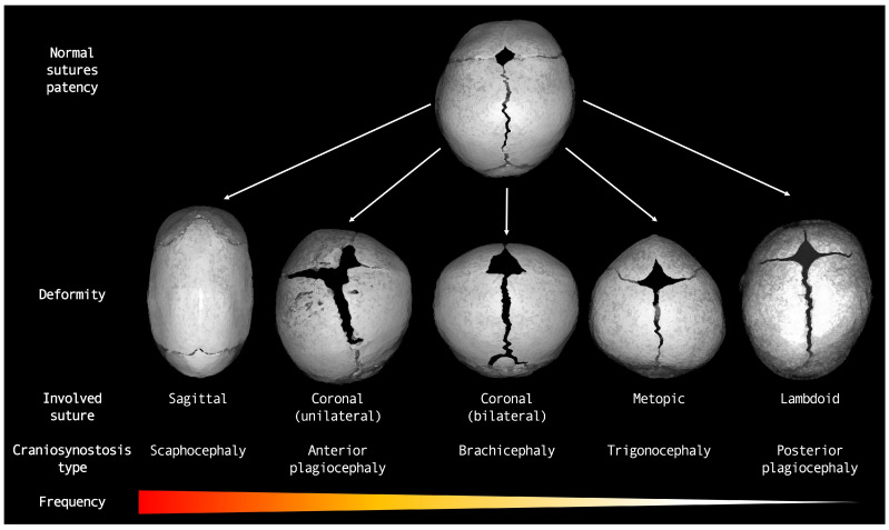
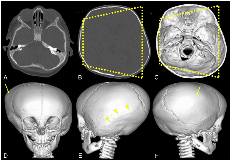
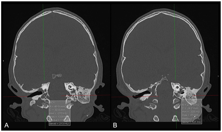
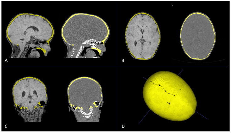
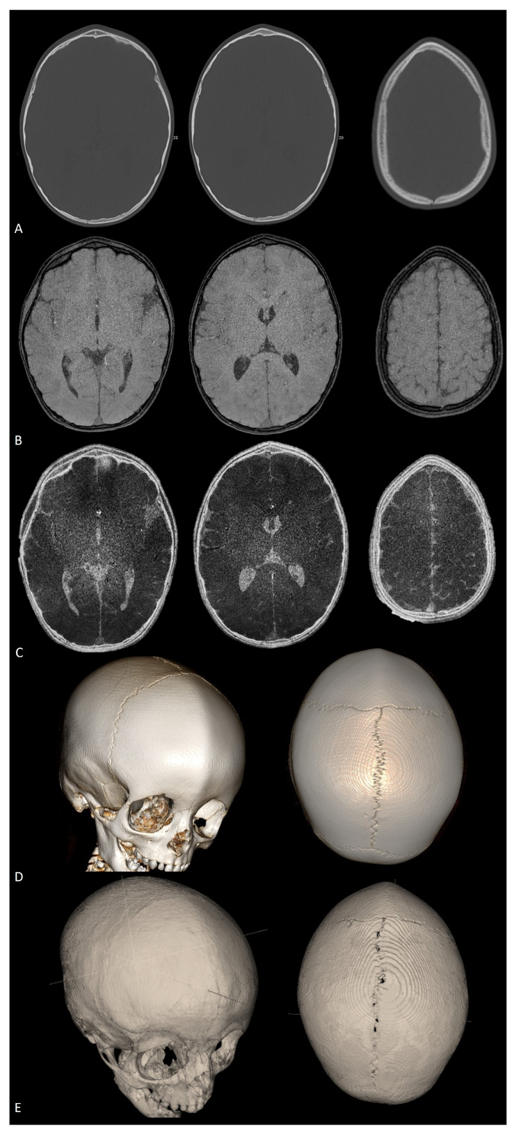
Craniosynostoses (CRS) are caused by the premature fusion of one or more cranial sutures, with isolated nonsyndromic CRS accounting for most of the clinical manifestations. Such premature suture fusion impacts both skull and brain morphology and involves regions far beyond the immediate area of fusion. The combined use of different neuroimaging tools allows for an accurate depiction of the most prominent clinical-radiological features in nonsyndromic CRS but can also contribute to a deeper investigation of more subtle alterations in the underlying nervous tissue organization that may impact normal brain development. This review paper aims to provide a comprehensive framework for a better understanding of the present and future potential applications of neuroimaging techniques for evaluating nonsyndromic CRS, highlighting strategies for optimizing their use in clinical practice and offering an overview of the most relevant technological advancements in terms of diagnostic performance, radiation exposure, and cost-effectiveness.

Keywords: arterial spin labeling; blackbone MRI; computed tomography; craniofacial surgery; craniosynostosis; magnetic resonance angiography; magnetic resonance imaging; neuroradiology; perfusion MRI.


Conflict of interest statement

The authors report no conflicts of interest concerning the materials or methods used in this study or the findings specified in this paper.

Figures

Figure 1
Figure 1
Diagram representing major types of nonsyndromic craniosynostoses.
Figure 2
Figure 2
A 3D CT image of a sagittal CRS (scaphocephaly) with an elongated skull, a prominent forehead, and occipital bulging. The white arrow indicates an abnormally fused suture.
Figure 3
Figure 3
A 3D CT image of a unilateral coronal CRS (anterior plagiocephaly) with an asymmetrical forehead, a flattened forehead and brow on the affected side, an elevated eye cavity on the affected side, and a nose that deviated toward the affected side. The white arrow indicates an abnormally fused suture.
Figure 4
Figure 4
A 3D CT example of bilateral coronal CRS (brachycephaly) with symmetrical flattening of the forehead, short broad skull, and increased ICP due to restricted growth. The white arrow indicates an abnormally fused suture.
Figure 5
Figure 5
A 3D CT example of metopic CRS (trigonocephaly) with a triangular keel-shaped forehead, hypotelorism, and a midline ridge along the forehead. The white arrow indicates an abnormally fused suture.
Figure 6
Figure 6
A 3D CT image of a lambdoid CRS (posterior plagiocephaly) with asymmetrical flattening of the back of the head, misalignment of the jaws, ears, and facial structures, and a tilted cranial base. The white arrow indicates an abnormally fused suture.
Figure 7
Figure 7
A 3D CT image of complex nonsyndromic CRS in a 6-month-old patient diagnosed with skull deformity. (A) Right frontal eminence asymmetry (white arrow) and minor left posterior plagiocephaly, with incipient sagittal synostosis (yellow arrow) and the presence of a right parietal accessory suture (red arrow). Subsequent 1-year follow-up examination (B) confirmed mild skull deformity due to complete fusion of the sagittal suture, not resulting in classic scaphocephaly due to patency of the PA suture.
Figure 8
Figure 8
Premature fusion of the sagittal suture in a 12-month-old boy. (A) CT volume rendering showing the dolichocephalic shape of the head, with complete fusion of the sagittal suture and sagittal ridge on the medial aspect of the suture. (B,C) US images obtained at the level of the yellow lines positioned on the 3D CT image showing the fused sagittal suture as a continuous hyperechoic calvaria with loss of the expected hypoechoic gap and the presence of a sagittal ridge (yellow arrow). (D) A small portion of the patient’s sagittal suture is shown on the very anterior aspect of the suture close to the anterior fontanelle as a hypoechoic gap (yellow arrowhead).
Figure 9
Figure 9
Example of high-resolution three-dimensional CT images (A,B) of the skull with volumetric image reconstruction (CF) in a patient diagnosed with posterior plagiocephaly due to right-sided lambdoid synostosis (yellow arrowheads), with a trapezoidal shape of the head (dotted yellow line) and compensatory left-sided frontal–parietal bossing (yellow arrow).
Figure 10
Figure 10
CT multiplanar coronal reconstruction of the jugular foramina in a patient diagnosed with brachycephaly showing a larger right jugular foramen ((A)—approximately 50 mm2) than on the left side ((B)—approximately 27 mm2) by means of area calculations obtained by placing manual regions of interest (ROIs—white dotted lines).
Figure 11
Figure 11
Multiplanar reconstruction (AC) of CT scan (left) and black-bone MR image (right) of a patient diagnosed with PT for premature fusion of the metopic suture. Axial (A), sagittal (B), and coronal (C) plane reconstructions were used to obtain bone segmentation (yellow) and subsequent volumetric rendering from black-bone MR images (D).
Figure 12
Figure 12
Axial CT scan (A), axial black-bone MRI (B), and axial black-bone MRI CT-like representation (C) at comparable levels in the same patient shown in Figure 11; 3D CT reconstruction (D) and 3D black-bone imaging postprocessing reconstruction (E) demonstrating comparable visualization of cranial sutures, metopic ridge, and overall skull deformity.
Figure 13
Figure 13
Non-contrast intracranial phase-contrast MR venography in a 28-month-old patient who underwent surgery for nonsyndromic complex CRS (premature fusion of lambdoid sutures, sagittal suture, and inferior aspect of left coronal suture), documenting progressive improvement in the visualization of dural venous sinuses (superior sagittal, transverse, straight, and sigmoid sinuses), larger deep cerebral veins (cavernous sinus), and cortical veins at three different time points: (A) before surgery; (B) 1 month after surgery; (C) 6 months after surgery.
Figure 14
Figure 14
Qualitative increase in cerebral blood flow demonstrated with ASL perfusion MRI before (upper row) and 6 months after surgery (bottom row) in three patients diagnosed with nonsyndromic isolated CRS: (A) scaphocephaly; (B) anterior plagiocephaly; and (C) trigonocephaly. Threshold color scale bars are shown on the left.
Figure 15
Figure 15
Axial black-bone MR image (A), axial TSE T2W image (B) and coronal TSE T2W image (C) of a 9-month-old female patient diagnosed with anterior plagiocephaly due to premature fusion of the right coronal suture (yellow arrows); aberrant adaptation of the right frontal lobe nervous tissue is indirectly demonstrated by the anomalous orientation of the frontal and frontal-insular sulci both on the axial and coronal planes (yellow arrowheads) compared to the left side, coupled to the asymmetric representation of adjacent CSF spaces (red asterisks). Axial FA maps (D) and color-coded representation of diffusion tensors on axial and coronal reconstructions superimposed on 3D-T1W images (E) confirmed altered fractional anisotropy (white dotted lines) and aberrant white matter fiber orientation (white dashed lines) due to white matter structural adaptive changes in the corresponding area. Color legend: red for left–right; blue for superior–inferior; and green for anterior–posterior.
Figure 16
Figure 16
Three cases of nonsyndromic trigonocephaly associated with (A) isolated tonsillar herniation with a normal ventricle size; (B) tonsillar herniation coupled to moderate supratentorial ventricular dilation; and (C) cervical spine syringomyelia.

References

    1. Johnson D., Wilkie A.O.M. Craniosynostosis. Eur. J. Hum. Genet. 2011;19:369–376. doi: 10.1038/ejhg.2010.235. - DOI - PMC - PubMed
    1. TBrah K., Thind R., Abel D.E. Craniosynostosis: Clinical Presentation, Genetics, and Prenatal Diagnosis. Obstet. Gynecol. Surv. 2020;75:636–644. doi: 10.1097/OGX.0000000000000830. - DOI - PubMed
    1. Blaser S.I., Padfield N., Chitayat D., Forrest C.R. Skull base development and craniosynostosis. Pediatr. Radiol. 2015;45:485–496. doi: 10.1007/s00247-015-3320-1. - DOI - PubMed
    1. Wilkie A.O., Johnson D., Wall S.A. Clinical genetics of craniosynostosis. Curr. Opin. Pediatr. 2017;29:622–628. doi: 10.1097/MOP.0000000000000542. - DOI - PMC - PubMed
    1. Passos-Bueno M.R., Sertié A.L., Jehee F.S., Fanganiello R., Yeh E. Genetics of Craniosynostosis: Genes, Syndromes, Mutations and Genotype-Phenotype Correlations. Front. Oral Biol. 2008;12:107–143. doi: 10.1159/000115035. - DOI - PubMed

LinkOut - more resources