. 2024 Aug 26;14(17):1868.
doi: 10.3390/diagnostics14171868.

The Impact of Liquid Biopsy in Advanced Ovarian Cancer Care

Affiliations

The Impact of Liquid Biopsy in Advanced Ovarian Cancer Care

Antoni Llueca et al. Diagnostics (Basel). .

Abstract

Introduction: Ovarian cancer is the third most common gynaecological cancer and has a very high mortality rate. The cornerstone of treatment is complete debulking surgery plus chemotherapy. Even with treatment, 80% of patients have a recurrence. Circulating tumour DNA (ctDNA) has been shown to be useful in the control and follow-up of some tumours. It could be an option to define complete cytoreduction and for the early diagnosis of recurrence.

Objective: We aimed to demonstrate the usefulness of ctDNA and cell-free DNA (cfDNA) as a marker of complete cytoreduction and during follow-up in patients with advanced ovarian cancer.

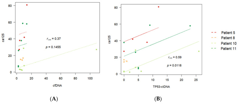
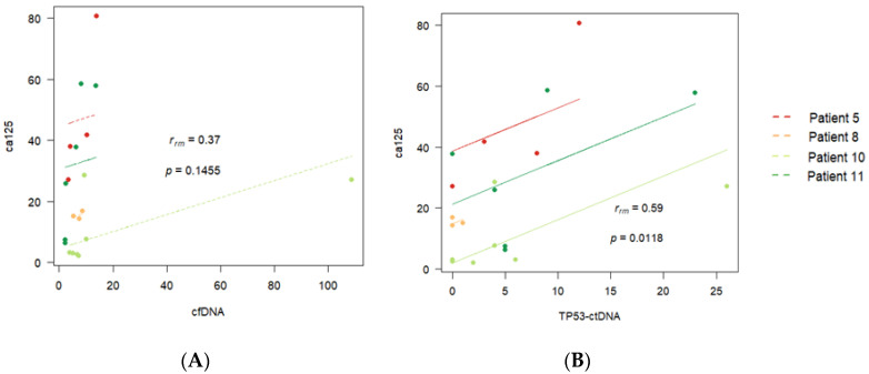
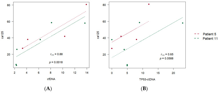
Material and methods: We selected 22 women diagnosed with advanced high-grade serous ovarian cancer, of which only 4 had complete records. We detected cfDNA by polymerase chain reaction (PCR), presented as ng/mL, and detected ctDNA with droplet digital PCR (ddPCR). We calculated Pearson correlation coefficients to evaluate correlations among cfDNA, ctDNA, and cancer antigen 125 (CA125), a biomarker.

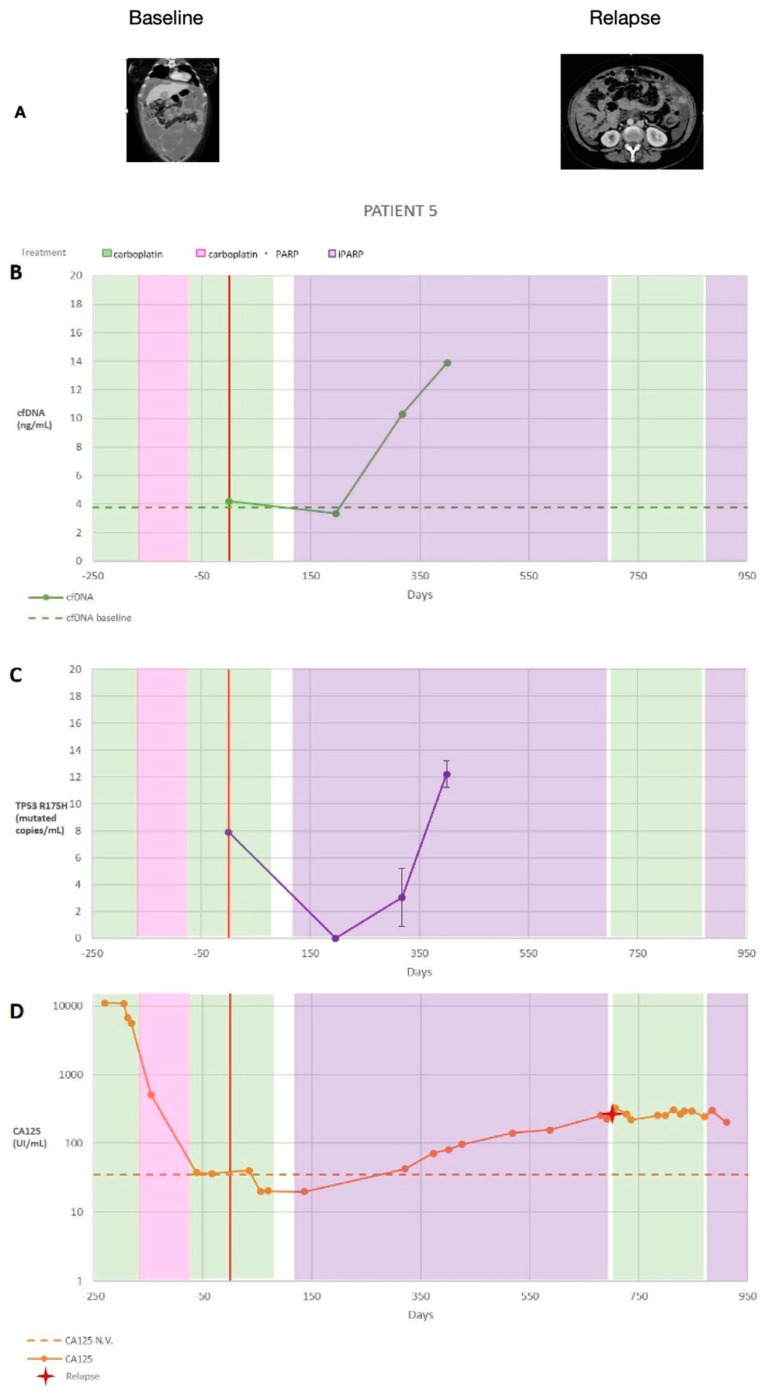
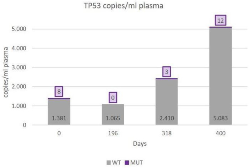
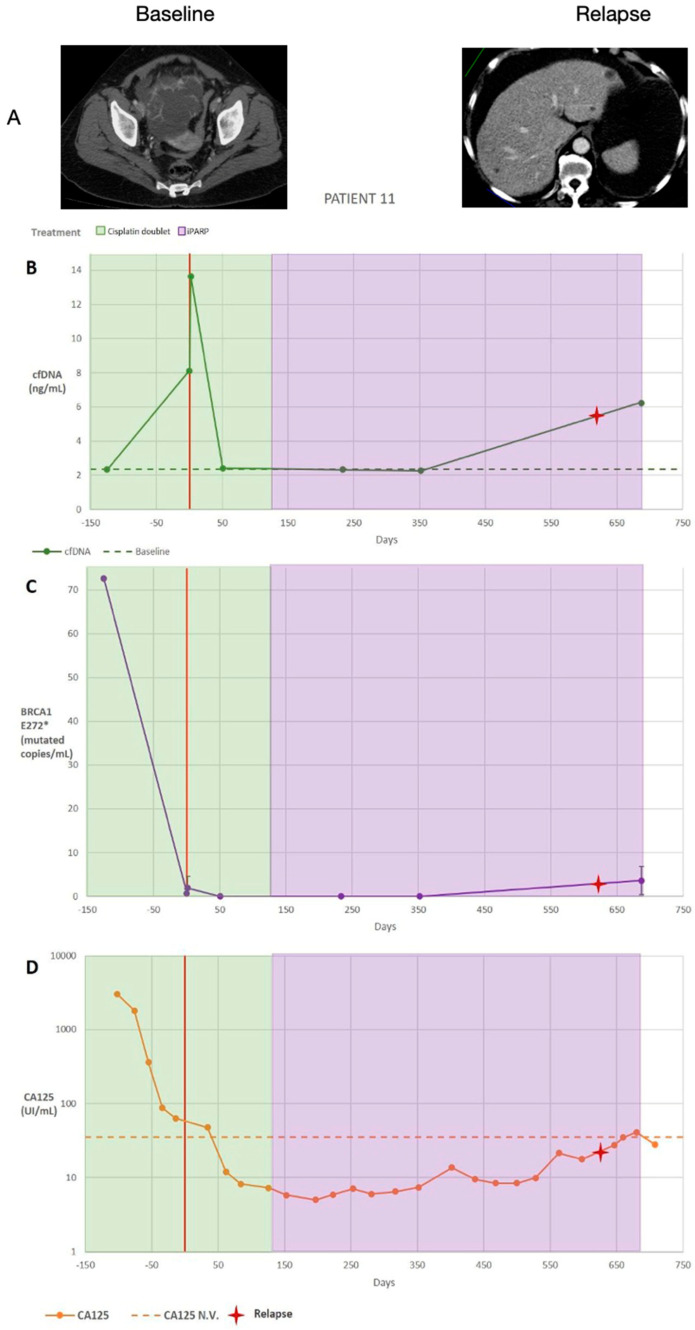
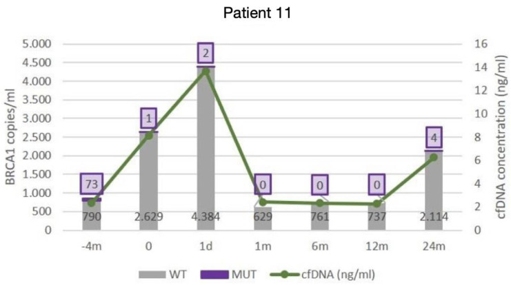
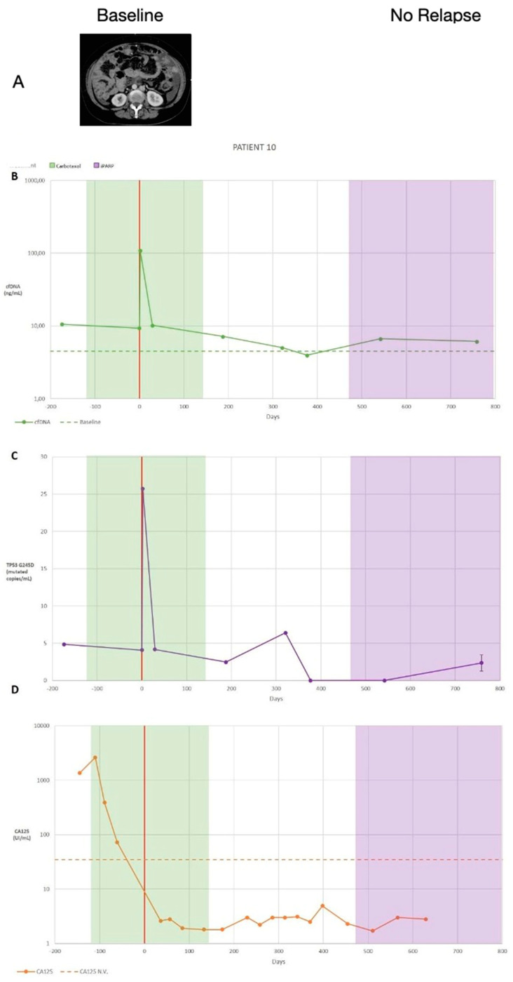
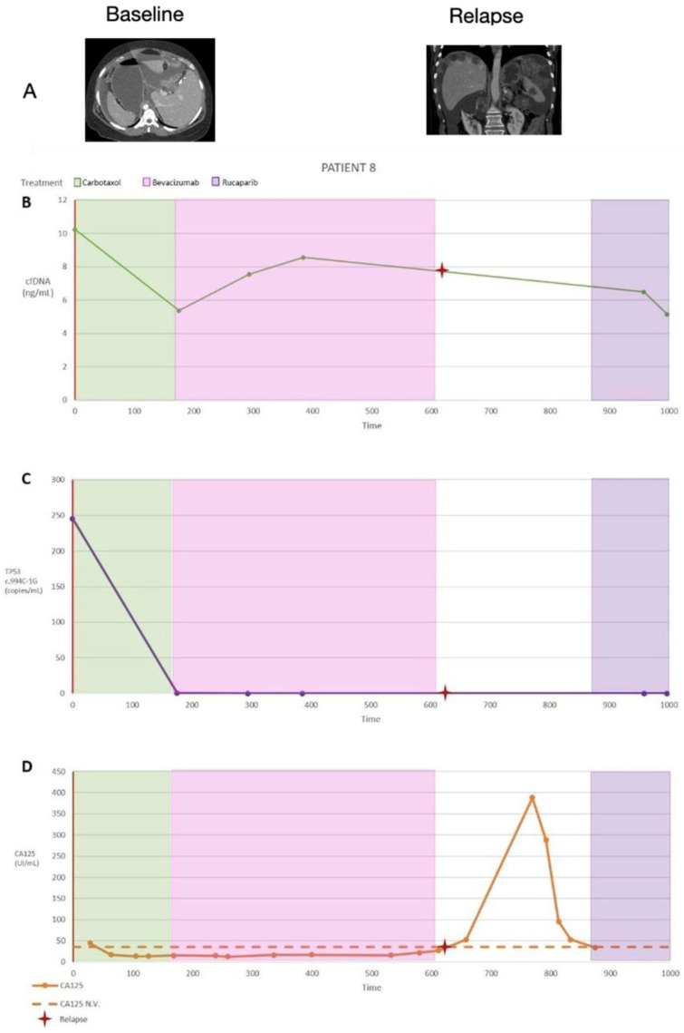
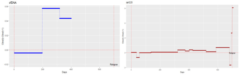
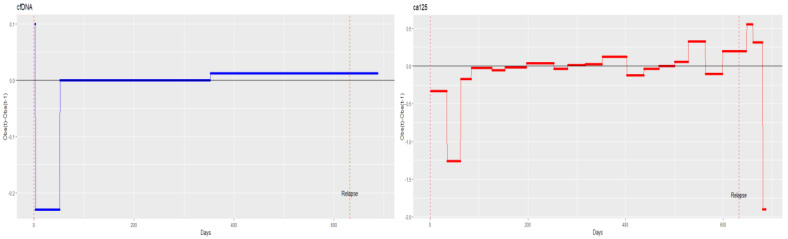
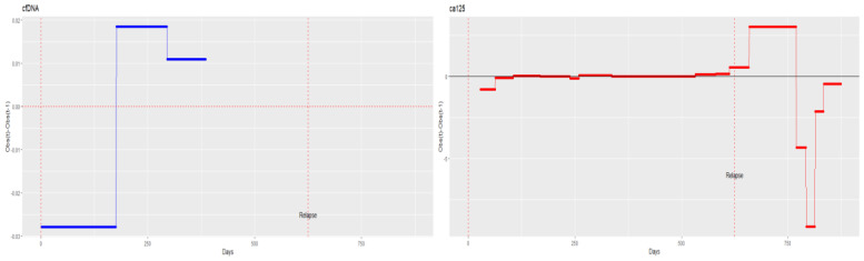
Results: The results obtained in the evaluation of cfDNA and ctDNA and their correlation with tumour markers and the radiology of patients with complete follow-up show disease progression during the disease, stable disease, or signs of recurrence. cfDNA and ctDNA correlated significantly with CA125. Following cfDNA and ctDNA over time indicated a recurrence several months earlier than computed tomography and CA125 changes.

Conclusion: An analysis of cfDNA and ctDNA offers a non-invasive clinical tool for monitoring the primary tumour to establish a complete cytoreduction and to diagnose recurrence early.

Keywords: advanced ovarian cancer; cancer care; liquid biopsy; translational research.


Conflict of interest statement

The authors declare no conflicts of interest.

Figures

Figure 1
Figure 1
(A) Summary of patient 5’s clinical information, including the treatment type and dates (treatment dates indicated by the coloured vertical stripes; the date of the cytoreduction surgery is indicated by a red vertical line, and the date of the diagnosed recurrence is indicated by a red cross) as well as computed tomography images and summarised results. (B) Cell-free DNA (cfDNA) plasma levels are presented as ng/mL. (C) Circulating tumour DNA (ctDNA) plasma levels are expressed as the number of mutated copies/mL of plasma. Replicates were used to quantify the average TP53 R175H copies for each time point, and the error bars represent the standard deviation across replicates; no bar indicates that the standard deviation was too low to be visualised on the scale used. The volume-adjusted limit of detection was 21.2 GEs (genome equivalent)/mL for all time points. A two-tailed t-test was used to compare the ctDNA levels between sequential time points. (D) Cancer antigen 125 (CA125) serum levels (U/mL); data from multiple replicates were not provided. * p = 0.05.
Figure 2
Figure 2
The number of TP53 (R175H) copies quantified by digital droplet polymerase chain reaction.
Figure 3
Figure 3
(A) Summary of patient 11’s clinical information, including the treatment type and dates (treatment dates indicated by the coloured vertical stripes; the date of the cytoreduction surgery is indicated by a red vertical line, and the date of a diagnosed relapse is indicated by a red cross) as well as computed tomography images and summarised results. (B) Cell-free DNA (cfDNA) plasma levels are presented as ng/mL. (C) Circulating tumour DNA (ctDNA) plasma levels are expressed as the number of mutated copies/mL. Replicates were used to quantify the average BRCA1 E272* (c.814 G>T) levels for each time point, and the error bars represent the standard deviation across replicates; no bars indicate that the standard deviation was too low to be visualised on the scale used. The volume-adjusted limit of detection was 21.2 GE/mL for all time points. A two-tailed t-test was used to compare the ctDNA quantities between sequential time points. (D) Cancer antigen 125 (CA125) serum levels (U/mL); data from multiple replicates were not provided. * p < 0.05, ** p < 0.01, and *** p < 0.001.
Figure 4
Figure 4
The number of BCRA1 E272* (c.814 G>T) copies quantified by digital droplet polymerase chain reaction.
Figure 5
Figure 5
(A) Summary of patient 10’s clinical information, including the treatment type and dates (treatment dates indicated by the coloured vertical stripes, and date of the cytoreduction surgery indicated by a red vertical line) as well as a computed tomography scan and summarised results. (B) Cell-free DNA (cfDNA) plasma levels are presented as ng/mL. (C) Circulating tumour DNA (ctDNA) plasma levels are expressed as the number of mutated copies/mL. Replicates were used to quantify the average TP53 G245 levels for each time point, and the error bars represent the standard deviation across replicates (n = 3); no bars indicate that the standard deviation was too low to be visualised on the scale used. The volume-adjusted limit of detection was 21.2 GE/mL for all time points. A two-tailed t-test was used to compare the ctDNA quantities between sequential time points. (D) Cancer antigen 125 (CA125) serum levels (U/mL); data from multiple replicates were not provided. * p < 0.05, ** p < 0.01, and *** p < 0.001.
Figure 6
Figure 6
(A) Summary of patient 8’s clinical information, including the treatment type and dates (treatment dates indicated by the coloured vertical stripes; the date of the cytoreduction surgery is indicated by a red vertical line, and the date of a diagnosed relapse is indicated by a red cross) as well as computed tomographic images and summarised results. (B) Cell-free DNA (cfDNA) plasma levels are presented as ng/mL. (C) Circulating tumour DNA (ctDNA) plasma levels are expressed as the number of mutated copies/mL. Replicates were used to quantify the average TP53 (c.994C-1G) levels for each time point, and the error bars represent the standard deviation across replicates (n = 3); no bars indicate that the standard deviation was too low to be visualised on the scale used. The volume-adjusted limit of detection was 21.2 GE/mL for all time points. A two-tailed t-test was used to compare the ctDNA quantities between sequential time points. (D) Cancer antigen 125 (CA125) serum levels (U/mL); data from multiple replicates were not provided. * p < 0.05, ** p < 0.01, and *** p < 0.001.
Figure 7
Figure 7
Aggregated correlation between (A) cell-free DNA (cfDNA) and CA125 and (B) TP53-ctDNA and CA125 for the four patients with complete records.
Figure 8
Figure 8
Aggregated correlation between (A) cfDNA and CA125 and (B) TP53-ctDNA and CA125 for eleven different patients of the dataset.
Figure 9
Figure 9
Aggregated correlation between (A) cfDNA and CA125 and (B) TP53 ctDNA and CA125 for two patients with the same disease progression on treatment.
Figure 10
Figure 10
Monitoring cell-free DNA (cfDNA) and cancer antigen 125 (CA125) over time in patient 5.
Figure 11
Figure 11
Monitoring cell-free DNA (cfDNA) and cancer antigen 125 (CA125) over time in patient 11.
Figure 12
Figure 12
Monitoring cell-free DNA (cfDNA) and cancer antigen 125 (CA125) over time in patient 8.

References

    1. Winter W.E., III, Maxwell G.L., Tian C., Carlson J.W., Ozols R.F., Rose P.G., Markman M., Armstrong D.K., Muggia F., McGuire W.P. Prognostic factors for stage III epithelial ovarian cancer: A Gynecologic Oncology Group Study. J. Clin. Oncol. 2007;25:3621–3627. doi: 10.1200/JCO.2006.10.2517. - DOI - PubMed
    1. Llueca A., Serra A., Maiocchi K., Delgado K., Jativa R., Gomez L., Escrig J. Predictive model for major complications after extensive abdominal surgery in primary advanced ovarian cancer. Int. J. Womens Health. 2019;11:161–167. doi: 10.2147/IJWH.S190493. - DOI - PMC - PubMed
    1. Llueca A., Serra A., Climent M.T., Segarra B., Maazouzi Y., Soriano M., Escrig J., on behalf MUAPOS Working Group Outcome quality standards in advanced ovarian cancer surgery. World J. Surg. Oncol. 2020;18:309. doi: 10.1186/s12957-020-02064-7. - DOI - PMC - PubMed
    1. Garzon S., Laganà A.S., Casarin J., Raffaelli R., Cromi A., Franchi M., Barra F., Alkatout I., Ferrero S., Ghezzi F. Secondary and tertiary ovarian cancer recurrence: What is the best management? Gland. Surg. 2020;9:1118–1129. doi: 10.21037/gs-20-325. - DOI - PMC - PubMed
    1. Climent M.T., Serra A., Llueca M., Llueca A. Surgery in Recurrent Ovarian Cancer: A Meta-Analysis. Cancers. 2023;15:3470. doi: 10.3390/cancers15133470. - DOI - PMC - PubMed

LinkOut - more resources