. 2024 Sep 9;13(17):1506.
doi: 10.3390/cells13171506.

Class Effect Unveiled: PPARγ Agonists and MEK Inhibitors in Cancer Cell Differentiation

Affiliations

Class Effect Unveiled: PPARγ Agonists and MEK Inhibitors in Cancer Cell Differentiation

Rakefet Ben-Yishay et al. Cells. .

Abstract

Epithelial-to-mesenchymal transition (EMT) plays a major role in breast cancer progression and the development of drug resistance. We have previously demonstrated a trans-differentiation therapeutic approach targeting invasive dedifferentiated cancer cells. Using a combination of PPARγ agonists and MEK inhibitors, we forced the differentiation of disseminating breast cancer cells into post-mitotic adipocytes. Utilizing murine breast cancer cells, we demonstrated a broad class effect of PPARγ agonists and MEK inhibitors in inducing cancer cell trans-differentiation into adipocytes. Both Rosiglitazone and Pioglitazone effectively induced adipogenesis in cancer cells, marked by PPARγ and C/EBPα upregulation, cytoskeleton rearrangement, and lipid droplet accumulation. All tested MEK inhibitors promoted adipogenesis in the presence of TGFβ, with Cobimetinib showing the most prominent effects. A metastasis ex vivo culture from a patient diagnosed with triple-negative breast cancer demonstrated a synergistic upregulation of PPARγ with the combination of Pioglitazone and Cobimetinib. Our results highlight the potential for new therapeutic strategies targeting cancer cell plasticity and the dedifferentiation phenotype in aggressive breast cancer subtypes. Combining differentiation treatments with standard therapeutic approaches may offer a strategy to overcome drug resistance.

Keywords: EMT; MEK inhibitor; PPARγ agonist; adipogenesis; breast cancer; differentiation therapy; thiazolidinediones.


Conflict of interest statement

Authors Vered Bar, Sara Aharon, Anna Kosenko and Adi Zundelevich were employed by the company Curesponse Ltd. The remaining authors declare that the research was conducted in the absence of any commercial or financial relationships that could be construed as a potential conflict of interest. The Curesponse Ltd. had no role in the design of the study; in the collection, analyses, or interpretation of data; in the writing of the manuscript, or in the decision to publish the results.

Figures
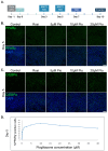
Figure 1
Figure 1
Pioglitazone upregulates adipogenesis-related transcription factors in murine breast cancer cells in a dose-dependent manner. (A) A scheme describing the optimized protocol to induce MTΔECad cells’ trans-differentiation into adipocytes with BMP2 and thiazolidinediones (TZDs). (B,C). MTΔECad cells were treated to induce adipogenesis as described in (A) for 5 days with BMP2 and either 2 μM Rosiglitazone (Rosi) or 5, 10, or 20μM Pioglitazone (Pio) as indicated. Control cells were treated with DMSO. Cells were then fixed and immunostained with antibodies against the adipogenesis-driving transcription factors PPARy (B) and C/EBPα (C) (top, green) and imaged with a confocal microscope. Merged images with DAPI nuclear counterstaining (blue) are presented in the bottom rows. Bars = 50 µm. (D) A graph demonstrating the percentage of PPARy-positive cells out of MTΔECad cells treated for adipogenesis with BMP2 and increasing concentrations of Pioglitazone for 5 days.
Figure 2
Figure 2
Pioglitazone induces trans-differentiation of murine breast cancer cells into well-differentiated adipocytes. (A) Adipogenesis was induced in MTΔECad cells for 10 days with 200 ng/mL BMP2 and either 2 μM Rosiglitazone (Rosi) or 10, 20, or 40 μM Pioglitazone (Pio) as indicated. Control cells were treated with DMSO. Cells were immunostained with anti C/EBPα antibody (green) and counterstained with Phalloidin to label F-actin (Magenta), visualizing actin rearrangement from stress fibers into cortical structures. Brightfield (BF) images demonstrate the formation of lipid droplets in the cytoplasm of treated cells. Bar = 100 µm. (B) Quantification of the percentage of C/EBPα-expressing cells in DMSO (control), BMP2 + Rosiglitazone (Rosi), or BMP2 + Pioglitazone (Pio) treated cells. After 10 days of cancer adipogenesis treatment, cells were fixed, immunostained with an anti C/EBPα antibody and DAPI to mark cell nuclei, and visualized with confocal microscopy. C/EBPα-positive cells were quantified n = 3. A two tailed t-test indicated no statistically significant difference between Rosi and Pio treatments (***, p < 0.001; N.S., p > 0.05). (C) MTΔECad Cells treated for 10 days with DMSO (control) or with BMP2 + Pioglitazone (Adipo) were immunostained with an antibody against the specific adipocyte marker Perilipin (a lipid droplet membrane protein, Magenta), and counterstained with BODIPY to visualize lipid droplets (green) and with DAPI (blue). Brightfield (BF) images of the same cells are shown on the right. Bar = 10 μm. (D) Representative images of MTΔECad cells treated for 10 days with DMSO (control), BMP2 + Rosiglitazone (Rosi), and BMP2 + Pioglitazone (Pio). On day 7 of adipogenesis treatment, cells were incubated with 5-ethynyl-20-deoxyuridine (EdU) for 72 hours to label proliferating cells (green). DAPI stain is shown in blue. Bar = 100 µm.
Figure 3
Figure 3
Class effect of MEK inhibitors enables cancer trans-differentiation in the presence of TGFβ (A) Schematic description of induction protocol for MTΔECad cells’ cancer adipogenesis in the presence of TGFβ with BMP2, thiazolidinediones (TZDs), and an MEK inhibitor (MEKi). (B) MTΔECad cells were treated as indicated in (A) with BMP2 + Pioglitazone (BMP2 + Pio), BMP2 + Pioglitazone and TGFβ, or BMP2 + Pioglitazone + TGFβ together with an MEK inhibitor (0.5 ng/mL Trametinib, 400 nM Binimetinib, 0.25 μM Selumetinib, or 0.25 μM Cobimetinib) as indicated. Control cells were treated with DMSO. After 10 days, cells were fixed, stained with BODIPY to mark lipid droplets (green) and with DAPI (blue), and imaged with a confocal microscope. DIC images of the same fields are shown at the bottom. Bar = 20 μm.
Figure 4
Figure 4
Pioglitazone and Cobimetinib can induce trans-differentiation of murine breast cancer cells into mature adipocytes in the presence of TGFβ. MTΔECad cells were treated for 10 days with BMP2 + Pioglitazone (BMP2 + Pio), BMP2 + Pioglitazone and TGFβ, or BMP2 + Pioglitazone + TGFβ together with the MEK inhibitor Cobimetinib. Cells were immunostained with antibodies against Perilipin (A) or PPARγ (B) (Magenta) and counterstained with BODIPY to mark lipid droplets (green) and DAPI (blue). Brightfield images of the same fields are shown in the bottom (BF). Bars = 20 µm.
Figure 5
Figure 5
C/EBPα is upregulated in cancer cells-induced adipogenesis regardless of TGFβ. Confocal imaging of MTΔECad cells treated for 10 days with BMP2 + Pioglitazone (BMP2 + Pio), BMP2 + Pioglitazone and TGFβ, or BMP2 + Pioglitazone + TGFβ together with Cobimetinib. Control cells were treated with DMSO. Cells were immunostained with anti-C/EBPα antibody (Magenta) and counterstained with BODIPY for lipid droplets labeling (green). DIC images are shown on the right. Bar = 100 µm.
Figure 6
Figure 6
Upregulation of PPARγ in ex vivo patient-derived breast cancer culture. (A) Three-dimensional tissues obtained from a core needle biopsy from the tumor of a triple-negative breast cancer patient were treated for 5 days with DMSO (control), Pioglitazone (Pio), Cobimetinib, or with a combination of Pioglitazone and Cobimetinib (Pio + Cobimetinib). Tissues were fixed and IHC staining with an antibody against PPARγ was performed (brown). Counterstaining with Hematoxylin is depicted in blue. Enlargement of the squared areas is shown on the right. Bar = 50 μm. (B) Quantification of the percentage of PPARy-positive cells in treated tissues.

References

    1. Haerinck J., Goossens S., Berx G. The epithelial–mesenchymal plasticity landscape: Principles of design and mechanisms of regulation. Nat. Rev. Genet. 2023;24:590–609. doi: 10.1038/s41576-023-00601-0. - DOI - PubMed
    1. Gerstberger S., Jiang Q., Ganesh K. Metastasis. Cell. 2023;186:1564–1579. doi: 10.1016/j.cell.2023.03.003. - DOI - PMC - PubMed
    1. Boumahdi S., de Sauvage F.J. The great escape: Tumour cell plasticity in resistance to targeted therapy. Nat. Rev. Drug Discov. 2019;19:39–56. doi: 10.1038/s41573-019-0044-1. - DOI - PubMed
    1. Nieto M.A. Epithelial Plasticity: A Common Theme in Embryonic and Cancer Cells. Science. 2013;342:1234850. doi: 10.1126/science.1234850. - DOI - PubMed
    1. Aiello N.M., Kang Y. Context-dependent EMT programs in cancer metastasis. J. Exp. Med. 2019;216:1016–1026. doi: 10.1084/jem.20181827. - DOI - PMC - PubMed

Publication types

MeSH terms

LinkOut - more resources