. 2024 Aug 23;25(17):9148.
doi: 10.3390/ijms25179148.

Effect of Silicon Nitride Coating on Titanium Surface: Biocompatibility and Antibacterial Properties

Affiliations

Effect of Silicon Nitride Coating on Titanium Surface: Biocompatibility and Antibacterial Properties

Akina Tani et al. Int J Mol Sci. .

Abstract

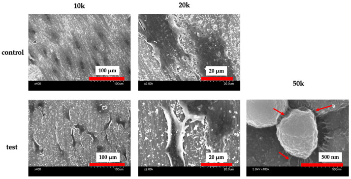
In recent years, with the advent of a super-aged society, lifelong dental care has gained increasing emphasis, and implant therapy for patients with an edentulous jaw has become a significant option. However, for implant therapy to be suitable for elderly patients with reduced regenerative and immunological capabilities, higher osteoconductive and antimicrobial properties are required on the implant surfaces. Silicon nitride, a non-oxide ceramic known for its excellent mechanical properties and biocompatibility, has demonstrated high potential for inducing hard tissue differentiation and exhibiting antibacterial properties. In this study, silicon nitride was deposited on pure titanium metal surfaces and evaluated for its biocompatibility and antibacterial properties. The findings indicate that silicon nitride improves the hydrophilicity of the material surface, enhancing the initial adhesion of rat bone marrow cells and promoting hard tissue differentiation. Additionally, the antibacterial properties were assessed using Staphylococcus aureus, revealing that the silicon nitride-coated surfaces exhibited significant antibacterial activity. Importantly, no cytotoxicity was observed, suggesting that silicon nitride-coated titanium could serve as a novel implant material.

Keywords: antibacterial material; biocompatibility; implant; silicon nitride; titanium.


Conflict of interest statement

The authors declare that there are no conflicts of interest.

Figures

Figure 1
Figure 1
SEM analysis of the control and test groups. Silicon nitride crystal deposition was observed on the surface of the material in the experimental group.
Figure 2
Figure 2
XPS analysis of the control and test groups (a). The XPS spectra of a wide scan of the test group (b). The presence of Si and N, the main elements of silicon nitride, was observed on the material surfaces of the experimental group, while Ti was not detected.
Figure 3
Figure 3
Contact angle analysis of the control and test groups (a). A decrease in contact angle was observed in the experimental group. The contact angle of the test group was lower than that of the control group (n = 4, ** p < 0.01) (b).
Figure 4
Figure 4
The results of the BSA adhesion examination showing significantly higher adsorption in the test group compared to the control group (n = 4, * p < 0.1, ** p < 0.01, *** p < 0.001, **** p < 0.0001).
Figure 5
Figure 5
Fluorescence microscope observations of RBMCs’ morphology on the titanium surface after 6 h of culture. RBMCs’ adhesion to the surface of the materials was confirmed for both groups.
Figure 6
Figure 6
After Si3N4 coating on the material surface, the number of adhered RBMCs was statistically significantly higher than that in the control group (n = 4, ** p < 0.01, *** p < 0.001).
Figure 7
Figure 7
Alkaline phosphatase (ALP) expression in bone marrow cells at days 7 and 14 after the start of the culture was significantly higher on the material surface of the test group compared to the control group (n = 4, ** p < 0.01).
Figure 8
Figure 8
Calcium deposition in bone marrow cells at days 21 and 28 after the start of the culture was significantly higher on the material surface of the test group compared to the control group (n = 4, ** p < 0.01).
Figure 9
Figure 9
Gene expression related to the induction of hard tissue differentiation was analyzed on the material surface of samples from the test and control groups. The assay was performed at specific measurement times for each gene. Significantly higher gene expression was observed on the material surface of the test group compared to the control group at all measurement time points (n = 4, * p < 0.1, ** p < 0.01).
Figure 10
Figure 10
SEM images of the material surfaces of the test and control groups seeded with Staphylococcus aureus are depicted. It is evident that there was minimal bacterial adhesion to the surface of the experimental group. Furthermore, at high magnification, bacteria were observed to be attached to the silicon nitride crystals via pseudopodia (red arrow).
Figure 11
Figure 11
The results of the bacterial adhesion test using Staphylococcus aureus biofilms are presented. Significantly lower values were observed in the test group compared to the control group (n = 4, **** p < 0.0001).
Figure 12
Figure 12
The evaluation of cytotoxicity using V79 cells. The material surface did not affect cell growth in both the test and control groups.

References

    1. Nanci A., Wuest J.D., Peru L., Brunet P., Sharma V., Zalzal S., McKee M.D. Chemical modification of titanium surfaces for covalent attachment of biological molecules. J. Biomed. Mater. Res. 1998;40:324–335. doi: 10.1002/(SICI)1097-4636(199805)40:2<324::AID-JBM18>3.0.CO;2-L. - DOI - PubMed
    1. Ogawa T., Nishimura I. Different bone integration profiles of turned and acid-etched implants associated with modulated expression of extracellular matrix genes. Int. J. Oral Maxillofac. Implant. 2003;18:200–210. - PubMed
    1. Wang T., Ouyang H., Luo Y., Xue J., Zheng L. Rehabilitation exercise–driven symbiotic electrical stimulation system accelerating bone regeneration. Sci. Adv. 2024;10:eadi6799. doi: 10.1126/sciadv.adi6799. - DOI - PMC - PubMed
    1. Vandrovcová M., Bačáková L. Adhesion, growth and differentiation of osteoblasts on surface-modified materials developed for bone implants. Physiol. Res. 2011;60:403–417. doi: 10.33549/physiolres.932045. - DOI - PubMed
    1. Mendonça G., Mendonça D.B.S., Aragão F.J.L., Cooper L.F. Advancing dental implant surface technology-From micron-to nanotopography. Biomaterials. 2008;29:3822–3835. doi: 10.1016/j.biomaterials.2008.05.012. - DOI - PubMed

MeSH terms

LinkOut - more resources