. 2024 Aug 23;25(17):9166.
doi: 10.3390/ijms25179166.

Novel Copper (II) Complexes with Fluorine-Containing Reduced Schiff Base Ligands Showing Marked Cytotoxicity in the HepG2 Cancer Cell Line

Affiliations

Novel Copper (II) Complexes with Fluorine-Containing Reduced Schiff Base Ligands Showing Marked Cytotoxicity in the HepG2 Cancer Cell Line

Bianka Oboňová et al. Int J Mol Sci. .

Abstract

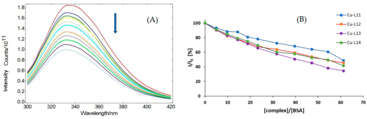
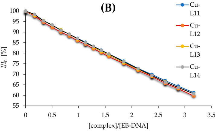
Several novel copper (II) complexes of reduced Schiff bases containing fluoride substituents were prepared and structurally characterized by single-crystal X-ray diffraction. The complexes exhibited diverse structures, with the central atom in distorted tetrahedral geometry. The biological effects of the products were evaluated, specifically their cytotoxicity, antimicrobial, and antiurease activities, as well as affinity for albumin (BSA) and DNA (ct-DNA). The complexes showed marked cytotoxic activities in the HepG2 hepatocellular carcinoma cell line, considerably higher than the standard cisplatin. The cytotoxicity depended significantly on the substitution pattern. The best activity was observed in the complex with a trifluoromethyl group in position 4 of the benzene ring-the dichloro[(±)-trans-N,N'-bis-(4-trifluoromethylbenzyl)-cyclohexane-1,2-diamine]copper (II) complex, whose activity (IC50 28.7 μM) was higher than that of the free ligand and markedly better than the activity of the standard cisplatin (IC50 336.8 μM). The same complex also showed the highest antimicrobial effect in vitro. The affinity of the complexes towards bovine serum albumin (BSA) and calf thymus DNA (ct-DNA) was established as well, indicating only marginal differences between the complexes. In addition, all complexes were shown to be excellent inhibitors of the enzyme urease, with the IC50 values in the lower micromolar region.

Keywords: BSA binding; DNA binding; anticancer activity; antimicrobial activity; copper; cytotoxicity; metal complexes; urease inhibition.


Conflict of interest statement

The authors declare no conflicts of interest.

Figures

Figure 1
Figure 1
Chemical structures of the Cu-L11Cu-L14 complexes.
Figure 2
Figure 2
Crystal structure of Cu-L11 (thermal ellipsoids shown at 30% probability level). The color scheme: gray/white—carbon and hydrogen, yellow—chlorine, green—fluorine, blue—nitrogen, purple—copper.
Figure 3
Figure 3
Crystal structure of Cu-L12 (thermal ellipsoids shown at 30% probability level). Solvent molecules and disordered atoms were omitted for the sake of clarity. The color scheme: gray/white—carbon and hydrogen, yellow—chlorine, green—fluorine, blue—nitrogen, purple—copper.
Figure 4
Figure 4
Crystal structure of Cu-L13 (thermal ellipsoids shown at 30% probability level). The color scheme: gray/white—carbon and hydrogen, yellow—chlorine, green—fluorine, blue—nitrogen, purple—copper.
Figure 5
Figure 5
Crystal structure of Cu-L14 (thermal ellipsoids shown at 30% probability level); organic part was set more transparent. Solvent molecules and disordered atoms were omitted for the sake of clarity. The color scheme: gray/white—carbon and hydrogen, yellow—chlorine, green—fluorine, blue—nitrogen, purple—copper.
Figure 6
Figure 6
(A) Representative fluorescence emission spectra of BSA in buffer solution in the presence of increasing concentrations of the complex Cu-L11. The arrow indicates the changes in fluorescence with increasing amounts of the complex. The binding of the copper complexes to albumin causes the induction of a conformational change in BSA and increased quenching of fluorescence in the tryptophane residues present in the molecule of BSA, leading to decreased fluorescence intensity. The concentration of added complexes ranged from 0 to 1.6 × 10⁻⁴ M (concentration increment 1.6 × 10⁻⁵ M, indicated by different color of the curve) (B) Graphical dependence of relative BSA fluorescence intensity in % at λ = 336 nm vs. concentration ratio [complex]/BSA for Cu-L11Cu-L14.
Figure 7
Figure 7
(A) Emission fluorescence spectra of EB-DNA in the buffer solution in the presence of increasing amount of complex Cu-L11. The arrow indicated the changes in fluorescence at increasing amounts of the complex. Insert graph shows the plot of I0/I vs. [complex]. The concentration of added complexes ranged from 0 to 2 × 10⁻⁴ M. (concentration increment 2 × 10⁻⁵ M, indicated by different color of the curve) (B) Plot of EB-DNA relative fluorescence intensity (I/I0, % at l = 614 nm vs. concentration ratio [complex]/[EB-DNA] for all tested complexes.
Figure 7
Figure 7
(A) Emission fluorescence spectra of EB-DNA in the buffer solution in the presence of increasing amount of complex Cu-L11. The arrow indicated the changes in fluorescence at increasing amounts of the complex. Insert graph shows the plot of I0/I vs. [complex]. The concentration of added complexes ranged from 0 to 2 × 10⁻⁴ M. (concentration increment 2 × 10⁻⁵ M, indicated by different color of the curve) (B) Plot of EB-DNA relative fluorescence intensity (I/I0, % at l = 614 nm vs. concentration ratio [complex]/[EB-DNA] for all tested complexes.
Figure 8
Figure 8
Cell viability (%) of the tested compounds in the concentration range of 20–250 μM. The values for the lowest measured concentrations (2–10 μM) were removed from this depiction for the sake of legibility, as the compounds are generally inefficient at these concentrations.

References

    1. Qin W., Long S., Panunzio M., Biondi S. Schiff bases: A short survey on an evergreen chemistry tool. Molecules. 2013;18:12264–12289. doi: 10.3390/molecules181012264. - DOI - PMC - PubMed
    1. Sankar R., Sharmila T.M. Schiff bases-based metallo complexes and their crucial role in the realm of pharmacology. A review. Results Chem. 2023;6:101179. doi: 10.1016/j.rechem.2023.101179. - DOI
    1. Nguyen Q.T., Jeong J.H. Syntheses and X-ray structures of Cu(II) and Zn(II) complexes of N,N′-dibenzyl-(R,R)-1,2-diaminocyclohexane and application to nitroaldol reaction. Polyhedron. 2008;27:3227–3230. doi: 10.1016/j.poly.2008.07.011. - DOI
    1. Sharma M., Joshi P., Kumar N., Joshi S., Rohilla R.K., Roy N., Rawat D.S. Synthesis, antimicrobial activity and structure-activity relationship study of N,N-dibenzyl-cyclohexane-1,2-diamine derivatives. Eur. J. Med. Chem. 2011;46:480–487. doi: 10.1016/j.ejmech.2010.11.027. - DOI - PubMed
    1. Rafii E., Dassonneville B., Heumann A. Highly selective R,S-coordination of non racemic (1R,2R)-(1,2-dialkyl)-1,2-diamine cyclohexane derivatives to palladium dichloride. Chem. Commun. 2007;2007:583–585. doi: 10.1039/b613961b. - DOI - PubMed

LinkOut - more resources