. 2024 Aug 24;25(17):9191.
doi: 10.3390/ijms25179191.

The Contribution of Mast Cells to the Regulation of Elastic Fiber Tensometry in the Skin Dermis of Children with Marfan Syndrome

Affiliations

The Contribution of Mast Cells to the Regulation of Elastic Fiber Tensometry in the Skin Dermis of Children with Marfan Syndrome

Dmitrii Atiakshin et al. Int J Mol Sci. .

Abstract

Marfan syndrome (MFS) is a hereditary condition accompanied by disorders in the structural and regulatory properties of connective tissue, including elastic fibers, due to a mutation in the gene encodes for fibrillin-1 protein (FBN1 gene) and the synthesis of abnormal fibrillin-1 glycoprotein. Despite the high potential of mast cells (MCs) to remodel the extracellular matrix (ECM), their pathogenetic significance in MFS has not been considered yet. The group of patients with Marfan syndrome included two mothers and five children (three girls aged 4, 11, and 11 and two boys aged 12 and 13). Normal skin was examined in two children aged 11 and 12. Histochemical, monoplex, and multiplex immunohistochemical techniques; combined protocols of simultaneous histochemical and immunohistochemical staining (the results of staining were assessed using light, epifluorescence, and confocal microscopy); and bioinformatics algorithms for the quantitative analysis of detected targets were used to evaluate mast cells and their relationship with other cells from extracellular structures in the skin dermis. Analysis of the skin MC population in children with Marfan syndrome revealed a considerably increased number of intra-organic populations with the preservation of the specific Tryptase+Chymase+CPA3+ protease profile typical of the skin. The features of the MC histotopography phenotype in MFS consisted of closer colocalization with elastic fibers, smooth muscle cells, and fibroblasts. MCs formed many intradermal clusters that synchronized the activity of cell functions in the stromal landscape of the tissue microenvironment with the help of spatial architectonics, including the formation of cell chains and the creation of fibrous niches. In MCs, the expression of specific proteases, TGF-β, and heparin increased, with targeted secretion of biologically active substances relative to the dermal elastic fibers, which had specific structural features in MFS, including abnormal variability in thickness along their entire length, alternating thickened and thinned areas, and uneven surface topography. This paper discusses the potential role of MCs in strain analysis (tensometry) of the tissue microenvironment in MFS. Thus, the quantitative and qualitative rearrangements of the skin MC population in MFS are aimed at altering the stromal landscape of the connective tissue. The results obtained should be taken into account when managing clinical signs of MFS manifested in other pathogenetically critical structures of internal organs, including the aorta, tendons, cartilage, and parenchymal organs.

Keywords: Marfan syndrome; carboxypeptidase A3; chymase; elastic fibers; heparin; mast cells; skin; tensometry extracellular matrix remodeling; tryptase.


Conflict of interest statement

The authors declare no conflicts of interest, and they confirm that this paper has not been and will not be published in whole or in part in any other journal and is not under consideration for publication elsewhere. This manuscript was approved by all authors.

Figures

Figure 1
Figure 1
Mast cell content in the skin of patients with Marfan syndrome (in %, relative content to other cells in the dermis). Notes: *—mother of patient No. 2; **—mother of patients No. 6 and No. 7; CPA3—Carboxypeptidase A3.
Figure 2
Figure 2
Histotopography of tryptase-positive mast cells in the skin dermis of patients with Marfan syndrome. Frequency of colocalization of MCs (%) with a fibrous component, αSMA-positive cells, and fibroblast. Staining technique: 1 multiplex detection of tryptase, elastic, and collagen fibers; 2 multiplex immunohistochemical detection of tryptase and α-SMA; 3 Giemsa staining. α-SMA: alpha-smooth muscle actin. Notes: *—mother of patient No. 2; **—mother of patients No. 6 and No. 7.
Figure 3
Figure 3
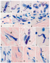
Skin mast cells in children without pathology. Technique: Giemsa staining. (a) Two contacting MCs with signs of secretion (arrowed). (b) MCs in the skin dermis: one of the cells has a long, narrow process (arrowed). (c,d) MC colocalization with fibroblasts (arrowed). (e) Adjacency of an MC to the postcapillary wall (arrowed). (f) A group of MCs in contact with each other and fibroblasts (arrowed). (g) MCs and a nuclear-free MC fragment located between bundles of collagen fibers (arrowed). (h,i) Elongated MCs in contact with fibrocytes (presumably arrowed) and collagen fibers (double arrowed). (j) MC that forms an outgrowth in the skin dermis. (k) An elongated MC adjacent to two fibroblasts. (l) A nuclear-free MC fragment among bundles of collagen fibers. (m) A degranulated MC with a predominantly peripheral localization of secretory granules. (n) MCs in contact with each other (arrowed), forming a functional chain throughout. Scale: 5 µm.
Figure 4
Figure 4
Mast cells in the fibrous landscape of the skin dermis in children without pathology. Technique: (aj) Weigert stain combined with tryptase immunohistochemical detection. (km) Weigert–Van Gieson stain combined with immunohistochemical tryptase detection. Notes: (a) general view of the elastic network in the papillary (arrowed) and reticular (double arrowed) layers of the skin dermis, with MCs. (b,c) MC colocalization with thin elastic fibers in the papillary layer of the skin dermis (arrowed). (hm) Interaction of MCs with elastic fibers in the reticular dermis. (h) MC in contact with several small-caliber (arrowed) and large-caliber (double arrowed) elastic fibers. (i,j) Tryptase-positive MC granules surrounding thick elastic fibers (arrowed). (k) Predominant localization of MC secretory granules in the area of elastic fibers. (l,m) Target tryptase secretion towards elastic fibers (arrowed) and collagen fibers (double arrowed). Scale: (a) 50 µm; the rest—5 µm.
Figure 5
Figure 5
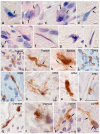
Mast cells in the skin dermis in Marfan syndrome. Technique: (ah) staining with Giemsa solution, (it) immunohistochemical staining for tryptase (il), carboxypeptidase A3 (mp), chymase (q,r), and CD117 (s,t). Notes: (ae) options for various MC colocalizations with fibroblasts (arrowed). (f) A nuclear-free area in the cytoplasm of an elongated MC. (g,h) Secretion of heparin-containing granules onto elastic fibers. (i) A group of MCs closely localized to fibroblasts. (j) Directed distribution of tryptase over long distances in the skin dermis (arrowed). (k) Active tryptase secretion onto the elastic fiber (arrowed). (l) Group of interacting CPA3+ MCs. (m) An elongated MC with a predominantly peripheral localization of CPA3 in the cytoplasm. (np) Active targeted secretion of CPA3 onto elastic fibers. MC granules adjacent to elastic fibers. (q,r) Active chymase degranulation onto fibers and cells of the skin dermis. (s,t) Predominantly peripheral localization of CD117 in the MC cytoplasm (arrowed). Scale: 5 µm. CPA3—Carboxypeptidase A3.
Figure 6
Figure 6
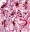
Target secretion of mast cell tryptase to the structural components of the elastic landscape of the skin dermis in Marfan syndrome. Technique: Combined Weigert staining with immunohistochemical detection of MC tryptase. Notes: (a,b) MC migration to areas with abnormal elastic fibers. (c) Intensive degranulation of MC with the development of elastic fiber degradation. (d) Spatial localization of MCs between three directions of elastic fibers. (eg) Targeted MC degranulation onto selected elastic fibers in the skin dermis (arrowed). (h) Accumulation of several MCs in areas of elastic fiber remodeling (arrowed). (im) Targeted tryptase secretion to the selective loci on elastic fibers of various thicknesses (arrowed). (n,o) Interaction of individual (n) or grouped (o) secretory granules (n) and the external component of elastic fibers from abnormal microfibrils with tryptase accumulation on the surface (arrowed). Scale: 5 µm.
Figure 7
Figure 7
Mast cell mapping in the fibrous landscape of the skin dermis in MFS. Technique: Combined Weigert–Van Gieson staining with immunohistochemical tryptase detection. Notes: (a) Simultaneous tryptase secretion onto the collagen and elastic fibers in the skin dermis (arrowed). (b) Formation of a pericellular zone of extracellular matrix remodeling (arrowed) and its impact on elastic fibers (double arrowed). (c) Active effect of tryptase on the collagen ECM (double arrowed) and extensive areas on the surface of elastic fibers (arrowed). (d) Selective localization of autonomous secretory MC granules in the elastic fiber area in the skin dermis (arrowed). (e) Selective tryptase secretion onto elastic fibers with a difference in their histochemical properties (arrowed). (f,g) Active entry of tryptase into areas of contact with collagen (arrowed) and elastic fibers (double arrowed). (h) Evident remodeling of the skin dermis by mast cells, with visible signs of elastic fiber destruction (indicated by an arrow). (i,j) Variants in MC localization in the tensometric areas of the elastic network of the skin dermis (arrowed), with intense tryptase secretion on the surface of the fibers. Scale: 5 µm.
Figure 8
Figure 8
Secretory profile of skin MCs in Marfan syndrome. Technique: Multiplex immunohistochemistry with simultaneous detection of tryptase, chymase, and CPA3 (a) and tryptase with αSMA (b,c) and TGFβ (d). Notes: (a) MCs in the skin dermis with a predominant Tryptase+Chymase+CPA3+ profile. (b,c) Colocalization of MCs with smooth myocytes in the microvasculature (double arrowed) and α-SMA-positive cells (arrowed), with active tryptase secretion. (d,e) High level of TGF-β expression in MCs in the skin dermis (arrowed). Scale: (c’) 2 µm; (e) 5 µm; the rest—10 µm. CPA3: carboxypeptidase A3. TGF-β: transforming growth factor beta.

References

    1. Pyeritz R.E. Recent progress in understanding the natural and clinical histories of the Marfan syndrome. Trends Cardiovasc. Med. 2016;26:423–428. doi: 10.1016/j.tcm.2015.12.003. - DOI - PubMed
    1. Wheeler J.B., Ikonomidis J.S., Jones J.A. Connective Tissue Disorders and Cardiovascular Complications: The Indomitable Role of Transforming Growth Factor-beta Signaling. Adv. Exp. Med. Biol. 2021;1348:161–184. doi: 10.1007/978-3-030-80614-9_7. - DOI - PMC - PubMed
    1. Kielty C.M. Fell-Muir Lecture: Fibrillin microfibrils: Structural tensometers of elastic tissues? Int. J. Exp. Pathol. 2017;98:172–190. doi: 10.1111/iep.12239. - DOI - PMC - PubMed
    1. Wagenseil J.E., Mecham R.P. New insights into elastic fiber assembly. Birth Defects Res. C Embryo Today. 2007;81:229–240. doi: 10.1002/bdrc.20111. - DOI - PubMed
    1. Godfrey M. From fluorescence to the gene: The skin in the Marfan syndrome. J. Investig. Dermatol. 1994;103:58S–62S. doi: 10.1038/jid.1994.11. - DOI - PubMed

MeSH terms

LinkOut - more resources