. 2024 Aug 26;25(17):9241.
doi: 10.3390/ijms25179241.

Synergistic Cytotoxicity of Histone Deacetylase and Poly-ADP Ribose Polymerase Inhibitors and Decitabine in Breast and Ovarian Cancer Cells: Implications for Novel Therapeutic Combinations

Affiliations

Synergistic Cytotoxicity of Histone Deacetylase and Poly-ADP Ribose Polymerase Inhibitors and Decitabine in Breast and Ovarian Cancer Cells: Implications for Novel Therapeutic Combinations

Benigno C Valdez et al. Int J Mol Sci. .

Abstract

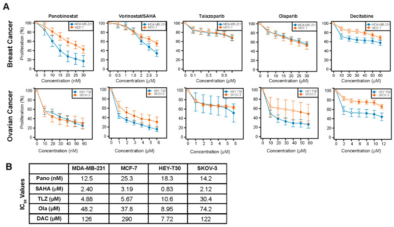
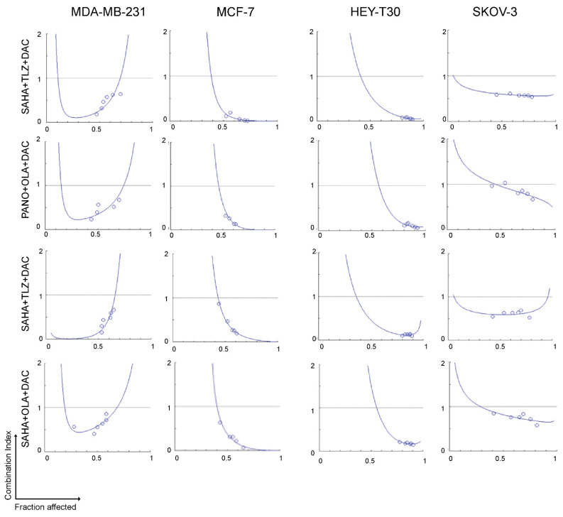
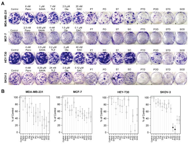
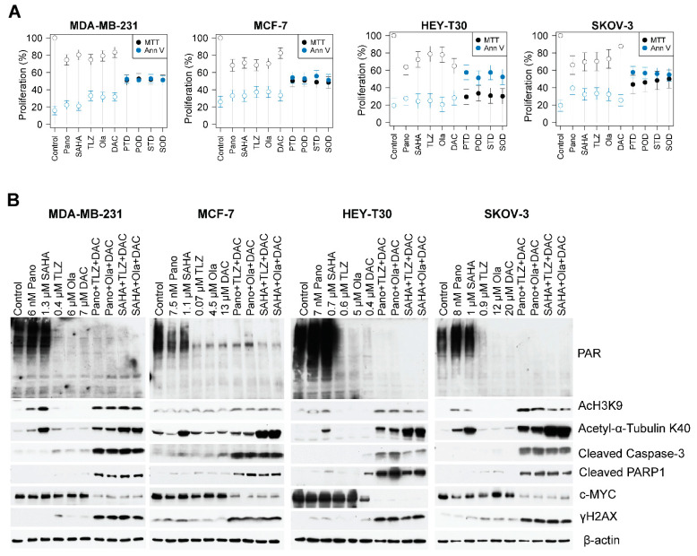
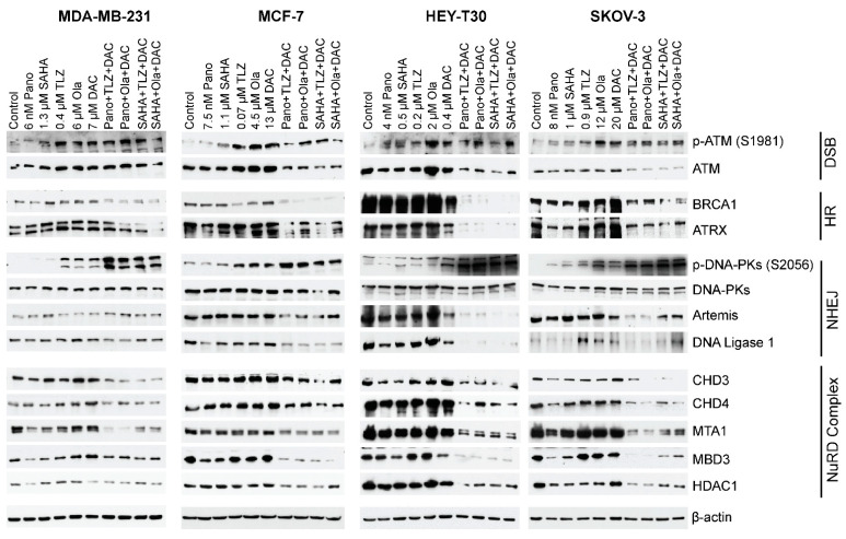
Breast and ovarian cancers pose significant therapeutic challenges. We explored the synergistic cytotoxicity of histone deacetylase inhibitors (HDACis), poly(ADP-ribose) polymerase inhibitors (PARPis), and decitabine in breast (MDA-MB-231 and MCF-7) and ovarian (HEY-T30 and SKOV-3) cancer cell lines that were exposed to HDACi (panobinostat or vorinostat), PARPi (talazoparib or olaparib), decitabine, or their combinations. HDACi, PARPi, and decitabine combinations had synergistic cytotoxicity (assessed by MTT and clonogenic assays) in all cell lines (combination index < 1). Clonogenic assays confirmed the sensitivity of breast and ovarian cancer cell lines to the three-drug combinations (panobinostat, talazoparib, and decitabine; panobinostat, olaparib, and decitabine; vorinostat, talazoparib, and decitabine; vorinostat, olaparib, and decitabine). Cell proliferation was inhibited by 48-70%, and Annexin V positivity was 42-59% in all cell lines exposed to the three-drug combinations. Western blot analysis showed protein PARylation inhibition, caspase 3 and PARP1 cleavage, and c-MYC down-regulation. The three-drug combinations induced more DNA damage (increased phosphorylation of histone 2AX) than the individual drugs, impaired the DNA repair pathways, and altered the epigenetic regulation of gene expression. These results indicate that HDACi, PARPi, and decitabine combinations should be further explored in these tumor types. Further clinical validation is warranted to assess their safety and efficacy.

Keywords: DNA repair; HDAC inhibitors; PARP inhibitors; breast cancer; decitabine; ovarian cancer; synergistic cytotoxicity.


Conflict of interest statement

Apostolia M. Tsimberidou declares receipt of clinical trial research funding (to The University of Texas MD Anderson Cancer Center) from Agenus, IMMATICS, Novocure, OBI Pharma, Parker Institute for Cancer Immunotherapy, Tachyon, Tempus, and Tvardi; fees for consulting or advisory roles for Avstera Therapeutics, Bioeclipse, BrYet, Diaccurate, Macrogenics, NEX-I, and VinceRx; and travel expenses from ASCO, Cancer Care Crossroads, GenomeWeb conference, and Precision Medicine World Conference. The remaining authors declare no relevant conflicts of interest.

Figures

Figure 1
Figure 1
Dose–response curves for the studied drugs in breast and ovarian cancer cell lines. Cells were seeded in 96-well plates overnight and exposed to different concentrations of individual drugs for 3 days as described in Methods. Rate of cell proliferation was determined relative to control by MTT assay (A,B). Model-adjusted means are shown with 95% confidence intervals for the non-zero doses modeled, and solid points indicate a significant difference from the first non-zero dose (Supplemental Table S1). Each cell line of each drug was modeled independently. IC50 values were determined using CalcuSyn 2.0 software. Pano: panobinostat; SAHA: vorinostat; TLZ: talazoparib; Ola: olaparib; DAC: decitabine.
Figure 2
Figure 2
Synergistic cytotoxicity of HDACi, PARPi, and decitabine. Cells were seeded in 96-well plates overnight and exposed to drugs individually, or in three-drug combinations at a constant concentration ratio, and cell proliferation was analyzed after 3 days. The relationships between the calculated combination indexes (Y-axis) and fractions affected (X-axis) are shown. A combination index < 1.0 indicates synergism. The graphs are representative of two independent experiments.. SAHA: vorinostat; TLZ: talazoparib; DAC: decitabine; PANO: panobinostat; OLA: olaparib.
Figure 3
Figure 3
Colony formation assay. Cells were seeded in six-well plates overnight and exposed to individual drugs or three-drug combinations for 1–2 weeks and stained as described in Methods (A). Colony formation is presented relative to control (B). For the double- and triple-drug combinations, the drug concentrations were the same as indicated in the single-drug concentrations. Model-adjusted means are shown with 95% confidence intervals, and solid points indicate a significant synergistic difference from all of the component drugs (see Supplemental Table S2). Each cell line of each drug was modeled independently. Number of replicates = 3; Pano/P: panobinostat; SAHA/S: vorinostat; TLZ/T: talazoparib; Ola/O: olaparib; DAC/D: decitabine.
Figure 4
Figure 4
Drug-mediated inhibition of cell proliferation and PARylation and effects on survival and apoptosis protein markers. Cells were seeded in T25 flasks overnight and exposed to individual drugs or three-drug combinations for 3 days, harvested, and analyzed for cell proliferation by MTT assay (A) and Western blotting (B). For the double- and triple-drug combinations, the drug concentrations were the same as indicated in the single-drug concentrations. Model-adjusted means are shown with 95% confidence intervals, and solid points indicate a significant synergistic difference from all of the component drugs (Supplemental Table S3). Each cell line of each drug was modeled independently. Number of replicates = 3; Pano/P: panobinostat; SAHA/S: vorinostat; TLZ/T: talazoparib; Ola/O: olaparib; DAC/D: decitabine.
Figure 5
Figure 5
Effects of drugs on the levels of various proteins involved in DNA repair/DNA damage response. Cells were exposed to the indicated drug concentrations for 3 days prior to analysis by Western blotting. For the double- and triple-drug combinations, the drug concentrations were the same as indicated in the single-drug concentrations. Number of replicates = 3; DSB: double-strand break; HR: homologous recombination; NHEJ: non-homologous DNA end-joining; NuRD: nucleosome remodeling and deacetylase. Pano: panobinostat; SAHA: vorinostat; TLZ: talazoparib; Ola: olaparib; DAC: decitabine.

References

    1. Domcke S., Sinha R., Levine D.A., Sander C., Schultz N. Evaluating cell lines as tumour models by comparison of genomic profiles. Nat. Commun. 2013;4:2126. doi: 10.1038/ncomms3126. - DOI - PMC - PubMed
    1. American Cancer Society Key Statistics for Breast Cancer. [(accessed on 29 January 2024)]. Available online: https://www.cancer.org/cancer/types/breast-cancer/about/how-common-is-br....
    1. American Cancer Society Key Statistics for Breast Cancer in Men. [(accessed on 29 January 2024)]. Available online: https://www.cancer.org/cancer/types/breast-cancer-in-men/about/key-stati....
    1. American Cancer Society Key Statistics for Ovarian Cancer. [(accessed on 29 January 2024)]. Available online: https://www.cancer.org/cancer/types/ovarian-cancer/about/key-statistics.....
    1. Matei D.E., Nephew K.P. Epigenetic therapies for chemoresensitization of epithelial ovarian cancer. Gynecol. Oncol. 2010;116:195–201. doi: 10.1016/j.ygyno.2009.09.043. - DOI - PMC - PubMed

MeSH terms