

Mitigating Cold Ischemic Injury: HTK, UW and IGL-2 Solution's Role in Enhancing Antioxidant Defence and Reducing Inflammation in Steatotic Livers
- PMID: 39273266
- PMCID: PMC11394993
- DOI: 10.3390/ijms25179318
Mitigating Cold Ischemic Injury: HTK, UW and IGL-2 Solution's Role in Enhancing Antioxidant Defence and Reducing Inflammation in Steatotic Livers
Abstract
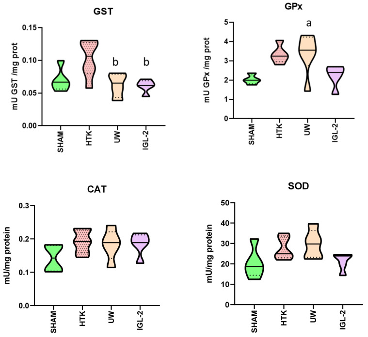
Liver transplantation remains the only definitive treatment for end-stage liver diseases. However, the increasing prevalence of fatty liver disease among potential donors exacerbates the shortage of suitable organs. This study evaluates the efficacy of the preservation solution Institut Georges Lopez-2 (IGL-2) compared to Histidine-Tryptophan-Ketoglutarate (HTK) and University of Wisconsin (UW) preservation solutions in mitigating ischemia-reperfusion injury (IRI) in steatotic livers. Using Zucker Obese rat livers, we assessed the impact of 24-h static cold storage (SCS) with each solution on transaminase release, glutathione redox balance, antioxidant enzyme activity, lipoperoxidation, and inflammation markers. IGL-2 and UW solutions demonstrated reduced transaminase and lactate levels compared to HTK, indicating better preservation of liver integrity. IGL-2 maintained a higher reduced glutathione/oxidized glutathione (GSH/GSSG) ratio, suggesting more effective management of oxidative stress. Antioxidant enzyme activities catalase, superoxide dismutase, and glutathione peroxidase (CAT, SOD, GPX) were higher in IGL-2 preserved livers, contributing to decreased oxidative damage. Lipid peroxidation markers and inflammatory markers were lower in IGL-2 than in HTK, indicating reduced oxidative stress and inflammation. Additionally, improved mitochondrial function was observed in the IGL-2 group, correlating with reduced reactive oxygen species (ROS) production and lipid peroxidation. These findings suggest that IGL-2 offers superior preservation of liver viability, reduces oxidative stress, and minimizes inflammation compared to HTK and UW solutions. By maintaining a higher ratio of reduced glutathione and antioxidant enzyme activity, IGL-2 effectively mitigates the harmful effects of ischemia-reperfusion injury. The reduced lipid peroxidation and inflammation in the IGL-2 group further underscore its potential in improving liver transplant outcomes. These results highlight the importance of optimizing preservation solutions to enhance the viability and functionality of donor organs, potentially expanding the donor pool and improving the success rates of liver transplantation. Future research should focus on refining preservation techniques and exploring additional protective agents to further improve organ preservation and transplant outcomes.
Keywords: HTK; IGL-2; UW; ischemic injury; oxidative stress; preservation solutions; static cold storage; steatotic liver; sterile inflammation.
Conflict of interest statement
The authors declare no conflicts of interest.
Figures


References
-
- Ghinolfi D., Melandro F., Torri F., Martinelli C., Cappello V., Babboni S., Silvestrini B., De Simone P., Basta G., Del Turco S. Extended criteria grafts and emerging therapeutics strategy in liver transplantation. The unstable balance between damage and repair. Transplant. Rev. 2021;35:100639. doi: 10.1016/j.trre.2021.100639. - DOI - PubMed
MeSH terms
Substances
Grants and funding
LinkOut - more resources
Full Text Sources
Medical
Research Materials
Miscellaneous
