Review
. 2024 Aug 30;25(17):9412.
doi: 10.3390/ijms25179412.

Expression of MicroRNAs in Adults with Celiac Disease: A Narrative Review

Affiliations
Review

Expression of MicroRNAs in Adults with Celiac Disease: A Narrative Review

Francielen Furieri Rigo et al. Int J Mol Sci. .

Abstract

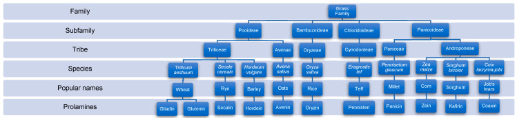
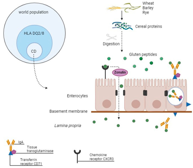
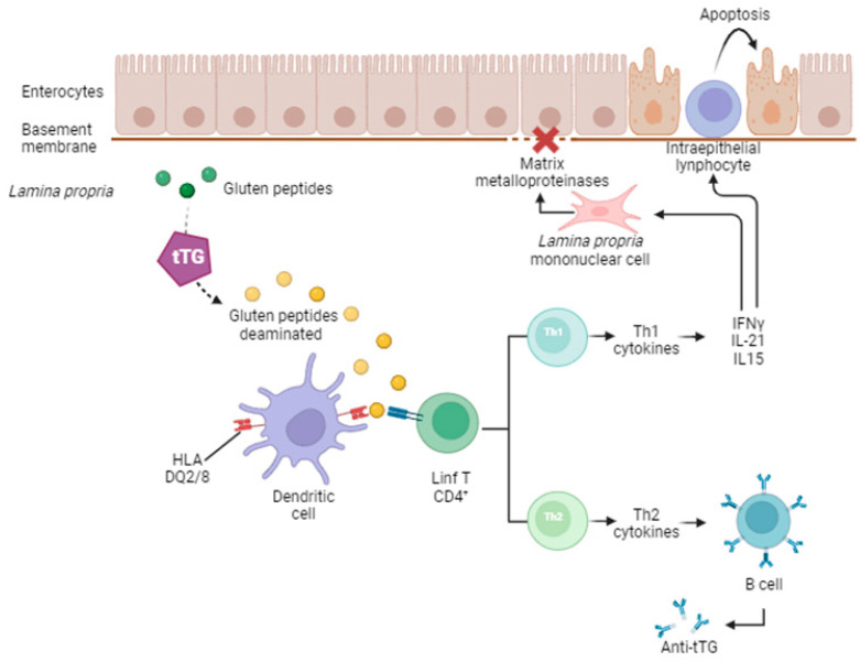
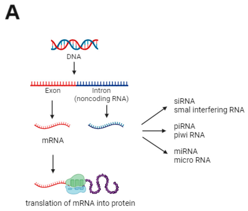
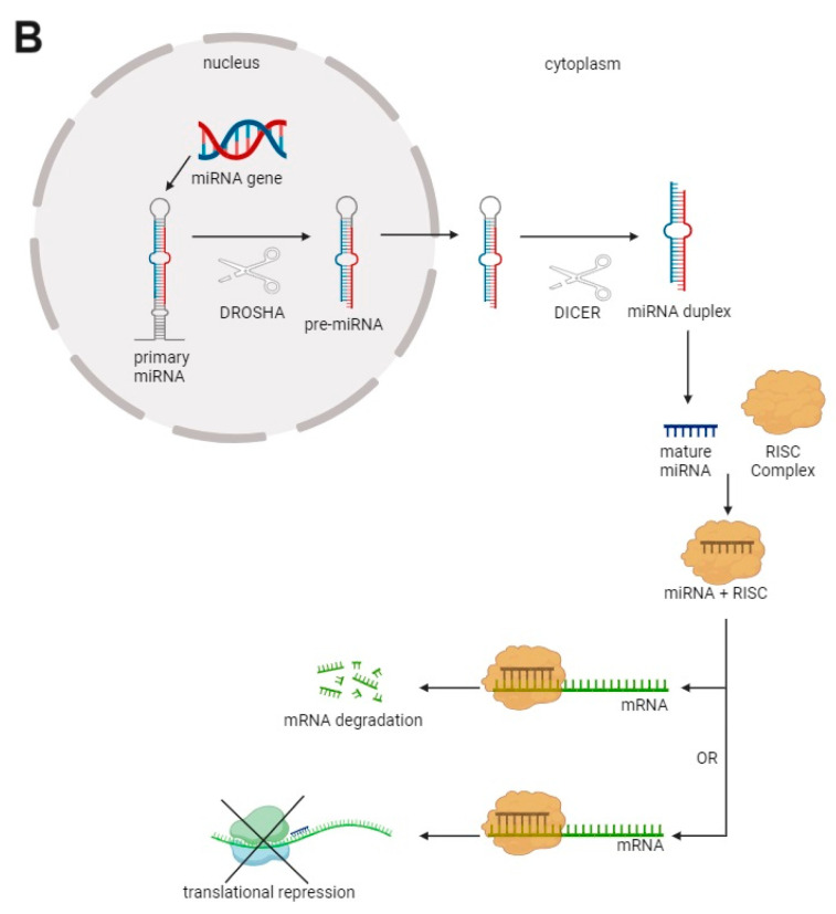
Celiac disease (CD) is an immune-mediated enteropathy triggered by the ingestion of proline- and glutamine-rich proteins, widely termed "gluten", in genetically susceptible individuals. CD induces an altered immune response that leads to chronic inflammation and duodenal mucosal damage. Currently, there are no specific tests for the accurate diagnosis of CD, and no drugs are available to treat this condition. The only available treatment strategy is lifelong adherence to a gluten-free diet. However, some studies have investigated the involvement of microRNAs (miRNAs) in CD pathogenesis. miRNAs are small noncoding ribonucleic acid molecules that regulate gene expression. Despite the growing number of studies on the role of miRNAs in autoimmune disorders, data on miRNAs and CD are scarce. Therefore, this study aimed to perform a literature review to summarize CD, miRNAs, and the potential interactions between miRNAs and CD in adults. This review shows that miRNA expression can suppress or stimulate pathways related to CD pathogenesis by regulating cell proliferation and differentiation, regulatory T-cell development, innate immune response, activation of the inflammatory cascade, focal adhesion, T-cell commitment, tissue transglutaminase synthesis, and cell cycle. Thus, identifying miRNAs and their related effects on CD could open new possibilities for diagnosis, prognosis, and follow-up of biomarkers.

Keywords: biomarkers; celiac disease; gluten-free diet; immune response; microRNAs.


Conflict of interest statement

The authors declare no conflicts of interest.

Figures

Figure 1
Figure 1
Phylogenetic relationships of some cereal species, names of family, subfamily, tribe, species, popular names, and their prolamines [25,26,27,28,29]. Flowchart generated using Microsoft Office 365 version 2408.
Figure 2
Figure 2
Simplified schematic representation of gluten peptide transport through the intestinal epithelium in patients with celiac disease. Up to 40% of healthy individuals carry human leukocyte antigen DQ2 or DQ8 haplotypes, but only 1% of the world population develops celiac disease (left diagram). During the digestion of wheat, barley, and rye, specific proteins called gluten release gluten peptides, which cross the basement membrane to the lamina propria in two ways: by the paracellular route and by retrotranscytosis. In the first mechanism, gluten peptides bind to the chemokine receptor CXCR3, which induces zonulin release with subsequent disassembly of tight junctions. In the second method, the peptides form a trimeric complex with secretory immunoglobulin A, transferrin receptor CD71, and tissue transglutaminase, which transfers the antigen from the apical to the basal side of the epithelium.
Figure 3
Figure 3
Simplified schematic representation of celiac disease pathogenesis. Once gluten peptides reach the lamina propria, they undergo deamination (dotted arrow) by tissue transglutaminase (tTG). Deaminated gluten peptides are rich in negatively charged glutamate residues, which increase their affinity and reinforce their presentation by dendritic cells to CD4+ T cells in the context of HLA-DQ2 or DQ8 molecules. Dendritic cells present deaminated gliadin peptides to CD4+ T cells. Activated gluten-reactive CD4+ T cells produce high levels of proinflammatory cytokines, including interferon (IFN)-γ, interleukin (IL)-21, and IL-15, thereby inducing T-helper (Th) cell type 1. This promotes inflammatory effects, including lamina propria mononuclear cell secretion of matrix metalloproteinases responsible for degradation of the extracellular matrix and basement membrane and increased cytotoxicity of CD8+ intraepithelial lymphocytes, as well as favoring apoptosis of lamina propria enterocytes. Furthermore, activated CD4+ T cells, through the production of Th2 cytokines, drive the activation and clonal expansion of B cells, resulting in the production of anti-tTG antibodies.
Figure 4
Figure 4
MicroRNA (miRNA) biogenesis and mode of action. (A) DNA contains regions called exons (nucleic acid coding sequences), which originate from messenger RNAs (mRNA) and are later transcribed into proteins, and introns (noncoding sequences), which are important to the origin of the miRNA; (B) inside the cell nucleus, miRNA is transcribed to primary miRNA and then to precursor miRNA (pre-miRNA) by the microprocessor complex Drosha/DGCR8 (DROSHA). The pre-miRNA is then transferred to the cell cytoplasm. Dicer participates in the second processing step to produce miRNA duplexes. The duplex is separated, and one strand is usually selected as the mature miRNA, whereas the other strand is degraded. The mature miRNA is loaded into an RNA-induced silencing complex (RISC) and regulates gene expression by binding to mRNA and causing its degradation or blocking its translation into proteins.
Figure 4
Figure 4
MicroRNA (miRNA) biogenesis and mode of action. (A) DNA contains regions called exons (nucleic acid coding sequences), which originate from messenger RNAs (mRNA) and are later transcribed into proteins, and introns (noncoding sequences), which are important to the origin of the miRNA; (B) inside the cell nucleus, miRNA is transcribed to primary miRNA and then to precursor miRNA (pre-miRNA) by the microprocessor complex Drosha/DGCR8 (DROSHA). The pre-miRNA is then transferred to the cell cytoplasm. Dicer participates in the second processing step to produce miRNA duplexes. The duplex is separated, and one strand is usually selected as the mature miRNA, whereas the other strand is degraded. The mature miRNA is loaded into an RNA-induced silencing complex (RISC) and regulates gene expression by binding to mRNA and causing its degradation or blocking its translation into proteins.

Similar articles

References

    1. Lebwohl B., Murray J.A., Verdú E.F., Crowe S.E., Dennis M., Fasano A., Green P.H.R., Guandalini S., Khosla C. Gluten introduction, breastfeeding, and celiac disease: Back to the drawing board. Am. J. Gastroenterol. 2016;111:12–14. doi: 10.1038/ajg.2015.219. - DOI - PMC - PubMed
    1. Felli C., Baldassarre A., Masotti A. Intestinal and circulating MicroRNAs in coeliac disease. Int. J. Mol. Sci. 2017;18:1907. doi: 10.3390/ijms18091907. - DOI - PMC - PubMed
    1. Caio G., Volta U., Sapone A., Leffler D.A., De Giorgio R., Catassi C., Fasano A. Celiac disease: A comprehensive current review. BMC Med. 2019;17:142. doi: 10.1186/s12916-019-1380-z. - DOI - PMC - PubMed
    1. Paolini A., Sarshar M., Felli C., Bruno S.P., Rostami-Nejad M., Ferretti F., Masotti A., Baldassarre A. Biomarkers to monitor adherence to gluten-free diet by celiac disease patients: Gluten immunogenic peptides and urinary miRNAs. Foods. 2022;11:1380. doi: 10.3390/foods11101380. - DOI - PMC - PubMed
    1. Rubio-Tapia A., Hill I.D., Semrad C., Kelly C.P., Greer K.B., Limketkai B.N., Lebwohl B. American College of Gastroenterology guidelines update: Diagnosis and management of celiac disease. Am. J. Gastroenterol. 2023;118:59–76. doi: 10.14309/ajg.0000000000002075. - DOI - PubMed

LinkOut - more resources