. 2024 Aug 30;25(17):9429.
doi: 10.3390/ijms25179429.

Advancing 3Rs: The Mouse Estrus Detector (MED) as a Low-Stress, Painless, and Efficient Tool for Estrus Determination in Mice

Affiliations

Advancing 3Rs: The Mouse Estrus Detector (MED) as a Low-Stress, Painless, and Efficient Tool for Estrus Determination in Mice

Irina V Belozertseva et al. Int J Mol Sci. .

Abstract

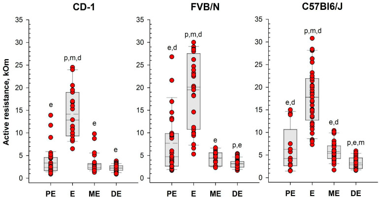
Determining the estrous cycle stages in mice is essential for optimizing breeding strategies, synchronizing experimental timelines, and facilitating studies in behavior, drug testing, and genetics. It is critical for reducing the production of genetically unmodified offspring in the generation and investigation of genetically modified animal models. An accurate detection of the estrus cycle is particularly relevant in the context of the 3Rs-Replacement, Reduction, and Refinement. The estrous cycle, encompassing the reproductive phases of mice, is key to refining experimental designs and addressing ethical issues related to the use of animals in research. This study presents results from two independent laboratories on the efficacy of the Mouse Estrus Detector (MED) from ELMI Ltd. (Latvia) for the accurate determination of the estrus phase. The female mice of five strains/stocks (CD1, FVB/N, C57Bl6/J, B6D2F1, and Swiss) were used. The results showed that the MEDProTM is a low-traumatic, simple, rapid, and painless method of estrus detection that supports the principles of the 3Rs. The use of the MEDProTM for estrus detection in mice caused minimal stress, enhanced mating efficiency, facilitated an increase in the number of embryos for in vitro fertilization, and allowed the production of the desired number of foster animals.

Keywords: 3Rs; female mice estrous cycle; mouse estrus detector; vaginal wall active resistance.


Conflict of interest statement

Dr. Dmitriy D. Merkulovs is co-inventor of patent LV15278B. The other co-authors have no conflict of interest to declare. ELMI Ltd. (Latvia) had no role in the design of the study; in the collection, analyses, or interpretation of data; in the writing of the manuscript; or in the decision to publish the results.

Figures

Figure 1
Figure 1
The proportion of female mice with a cytologically estimated estrus with a closed (C) or open (O) vagina: CD-1 (n = 71 and n = 39 for C and O, respectively); FVB/N (n = 79 and n = 31 for C and O, respectively); C57Bl6/J (n = 88 and n = 22 for C and O, respectively). *—p < 0.05 (Chi-square test).
Figure 2
Figure 2
Photo of the mouse estrus detector MEDProTM device (left panel) and a magnified image of its probe (right panel).
Figure 3
Figure 3
Measurement of vaginal wall active resistance in female mice by using the MEDProTM device.
Figure 4
Figure 4
Vaginal active resistance values as a function of the stage of the estrous cycle (PE—proestrus, E—estrus, ME—metestrus, DE—diestrus), determined by vaginal smear in female mice of different strains. Single-value plots show each observation’s value. The box represents the 25th and 75th percentiles and illustrates the median (solid line) and the mean (dashed line). The whiskers show the 10th and 90th percentiles. Letters indicate a significant difference (p < 0.05, Bonferroni test for multiple comparisons) as follows: p—proestrus, e—estrus, m—metestrus, d—diestrus.
Figure 5
Figure 5
Proportion of female mice with cytologically estimated estrus with AR < 6 kOm or AR > 6 kOm: CD-1 (n = 88 and n = 22, respectively); FVB/N (n = 68 and n = 42, respectively); C57Bl6/J (n = 55 and n = 55). *—p < 0.001 (Chi-square test).
Figure 6
Figure 6
Mating efficiency in female mice with different values of vaginal active resistance after overnight pairing with male. (left panel)—proportion of CD-1 females with vaginal plug (n = 97 for AR < 6 kOm; n = 25 for AR > 6 kOm); (right panel)—proportion of Swiss females that gave birth (n = 10 for ‘unknown”; n = 9 for AR > 6 kOm). *—p < 0.001 (Chi-square test); #—p < 0.001 (Fisher’s exact test).
Figure 7
Figure 7
Number of oocytes per mouse produced after mouse superovulation started at different vaginal AR values—AR < 6 kOm or AR > 6 kOm: FVB/N (n = 19 and n = 5, respectively); C57Bl6/J (n = 29 and n = 15, respectively); B6D2F1 (n = 51 and n = 9, respectively).
Figure 8
Figure 8
Locomotor activity and “emotionality” of Swiss female mice. A box-plot and whisker graph show the difference between the three experimental groups: (1) intact female mice (n = 21); (2) female mice 30 min after detector probe insertion into the vagina for measurement of active resistance (n = 21), and (3) female mice 30 min after vaginal lavage (n = 14). The box represents the 25th and 75th percentiles, and illustrates the median (solid line) and the mean (dashed line). The whiskers show the 10th and 90th percentiles and outliers are indicated as dots. * Indicate a significant difference (p < 0.05, Dunn’s test for multiple comparisons) from intact females.
Figure 9
Figure 9
Representative smears illustrating cytology for each estrous cycle stage: (A) proestrus; (B) estrus; (C) metestrus; (D) diestrus. Three cell types are identified: leukocytes (L), cornified epithelial cells (C), and nucleated epithelial cells (N).

Similar articles

References

    1. Dominguez-Oliva A., Hernandez-Avalos I., Martinez-Burnes J., Olmos-Hernandez A., Verduzco-Mendoza A., Mota-Rojas D. The Importance of Animal Models in Biomedical Research: Current Insights and Applications. Animals. 2023;13:1223. doi: 10.3390/ani13071223. - DOI - PMC - PubMed
    1. Liao B.Y., Zhang J. Low rates of expression profile divergence in highly expressed genes and tissue-specific genes during mammalian evolution. Mol. Biol. Evol. 2006;23:1119–1128. doi: 10.1093/molbev/msj119. - DOI - PubMed
    1. Monaco G., van Dam S., Casal Novo Ribeiro J.L., Larbi A., de Magalhaes J.P. A comparison of human and mouse gene co-expression networks reveals conservation and divergence at the tissue, pathway and disease levels. BMC Evol. Biol. 2015;15:259. doi: 10.1186/s12862-015-0534-7. - DOI - PMC - PubMed
    1. Miyagi A., Lu A., Humphreys B.D. Gene Editing: Powerful New Tools for Nephrology Research and Therapy. J. Am. Soc. Nephrol. 2016;27:2940–2947. doi: 10.1681/ASN.2016020146. - DOI - PMC - PubMed
    1. Mou H., Kennedy Z., Anderson D.G., Yin H., Xue W. Precision cancer mouse models through genome editing with CRISPR-Cas9. Genome Med. 2015;7:53. doi: 10.1186/s13073-015-0178-7. - DOI - PMC - PubMed

LinkOut - more resources