Review
. 2024 Aug 30;25(17):9455.
doi: 10.3390/ijms25179455.

Functions and Clinical Applications of Extracellular Vesicles in TH2 Cell-Mediated Airway Inflammatory Diseases: A Review

Affiliations
Review

Functions and Clinical Applications of Extracellular Vesicles in TH2 Cell-Mediated Airway Inflammatory Diseases: A Review

Jaehwan Cheon et al. Int J Mol Sci. .

Abstract

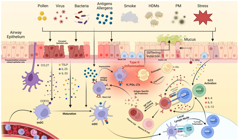
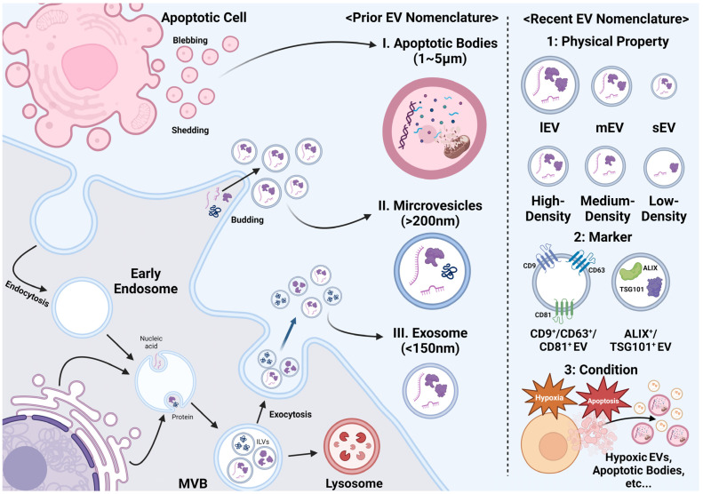
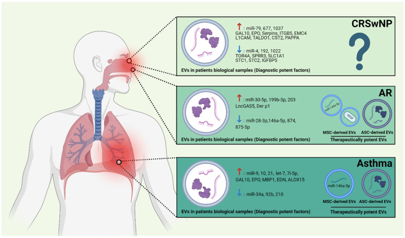
Type 2 airway inflammation (T2AI), driven by type 2 innate lymphoid and CD4+ T helper 2 cells, leads to various diseases and conditions, such as chronic rhinosinusitis with nasal polyps, allergic rhinitis, and asthma. Emerging evidence suggests the involvement of extracellular vesicles (EVs) in these diseases. In this review, we describe the immunological T2AI pathogenic mechanisms, outline EV characteristics, and highlight their applications in the diagnosis and treatment of T2AI. An extensive literature search was conducted using appropriate strategies to identify relevant articles from various online databases. EVs in various biological samples showed disease-specific characteristics for chronic rhinosinusitis with nasal polyps, allergic rhinitis, and asthma, with some demonstrating therapeutic effects against these conditions. However, most studies have been limited to in vitro and animal models, highlighting the need for further clinical research on the diagnostic and therapeutic applications of EVs.

Keywords: allergic rhinitis; asthma; chronic rhinosinusitis with nasal polyp; extracellular vesicles; type 2 inflammation.


Conflict of interest statement

The authors declare no conflicts of interest.

Figures

Figure 1
Figure 1
The pathogenesis of type 2 airway inflammation (T2AI). Various triggers, including pollens, viruses, bacteria, antigens/allergens, smoke, house dust mites (HDMs), particulate matter (PM), and stress, disrupt the airway epithelium, leading to the release of chemokines, such as the C-C motif chemokine ligand (CCL)-27, and alarmin cytokines, such as thymic stromal lymphopoietin (TSLP), interleukin (IL)-25, and IL-33. These signals activate type 2 innate lymphoid cells (ILC2s) and attract dendritic cells (DCs) expressing chemokine receptors, such as the C-C motif chemokine receptor (CCR)-10, to the site of disruption. Immature DCs (imDCs) migrate to the disrupted epithelium and are converted to mature DCs (mDCs) via stimulation by alarmin cytokines and interactions with the infected antigens/allergens. These mDCs present the antigens to naïve T-cells in local lymph nodes, promoting their polarization into TH2 cells. These TH2 cells, along with ILC2s, secrete cytokines (IL-4, IL-5, and IL-13), which influence various cell types, such as epithelial cells and eosinophils. Additionally, IL-4 and IL-13 facilitate B-cell class switching to IgE. Plasma cells secrete IgE, which binds to the IgE receptors (FcεR) on mast cell membranes via a process known as sensitization. Upon re-exposure to the antigen or allergen, the cross-linking of IgE on mast cells induces their degranulation, releasing inflammatory mediators, such as histamines (Hs), prostaglandins (PGs), and leukotrienes (LTs). Additionally, IL-5 activates and degranulates eosinophils. This integrative degranulation of mast cells and eosinophils ultimately leads to inflammatory symptoms in the airway. HDM, house dust mite; PM, particulate matter; CCL, C-C motif chemokine ligand; TSLP, thymic stromal lymphopoietin; IL, interleukin; ILC2, type 2 innate lymphoid cell; DC, dendritic cell; CCR, C-C motif chemokine receptor; imDC, immature DC; mDC, mature DC; Ig, immunoglobulin; FcεR, IgE receptor; H, histamine; PG, prostaglandin; LT, leukotriene.
Figure 2
Figure 2
The biogenesis and Classification of extracellular vesicles (EVs). EVs are classified into apoptotic bodies (ApoBDs), micro-vesicles, and exosomes based on their origin and size. ApoBDs are primarily formed via the shedding and blebbing of apoptotic cells, whereas micro-vesicles are produced via cellular budding. Exosomes are typically formed and released through the endosomal sorting complex required for the transport (ESCRT)-dependent pathway, with markers, such as tetraspanins, the tumor susceptibility gene 101 (TSG101), and the ALG-2-interacting protein X (ALIX), playing key roles in their identification. Recently, EV classification has become more nuanced, taking into account their size, density, biochemical composition, and condition of parent cells. EV, extracellular vesicle; ApoBD; apoptotic body; ESCRT, endosomal sorting complex required for transport; TSG, tumor susceptibility gene; ALIX, ALG-2-interacting protein X; lEV, large EV; mEV, medium EV; sEV, small EV.
Figure 3
Figure 3
Comprehensive applications of EVs for various T2AI diseases. EVs exhibit differentially expressed molecules in chronic rhinosinusitis with nasal polyps (CRSwNP), allergic rhinitis (AR), and asthma and can be applied for the diagnosis of these conditions. Specifically, mesenchymal stromal cells (MSCs) and adipose tissue-derived stem cells (ADSCs)-derived EVs are potential therapeutic targets for AR and asthma. ↑: Up-regulation, ↓: Down-regulation.

Similar articles

References

    1. Samitas K., Carter A., Kariyawasam H.H., Xanthou G. Upper and Lower Airway Remodelling Mechanisms in Asthma, Allergic Rhinitis and Chronic Rhinosinusitis: The One Airway Concept Revisited. Allergy. 2018;73:993–1002. doi: 10.1111/all.13373. - DOI - PubMed
    1. Fokkens W.J., Lund V.J., Hopkins C., Hellings P.W., Kern R., Reitsma S., Toppila-Salmi S., Bernal-Sprekelsen M., Alobid J.M.I., Anselmo-Lima Terezinha W., et al. European Position Paper on Rhinosinusitis and Nasal Polyps 2020. Rhinology. 2020;58((Suppl. S29)):1–464. doi: 10.4193/Rhin20.401. - DOI - PubMed
    1. Alnahas S., Abouammoh N., Althagafi W., Abd-Ellatif E.E. Prevalence, Severity, and Risk Factors of Allergic Rhinitis among Schoolchildren in Saudi Arabia: A National Cross-Sectional Study, 2019. World Allergy Organ. J. 2023;16:100824. doi: 10.1016/j.waojou.2023.100824. - DOI - PMC - PubMed
    1. Reddel H.K., Bacharier L.B., Bateman E.D., Brightling C.E., Brusselle G.G., Buhl R., Cruz A.A., Duijts L., Drazen J.M., FitzGerald J.M., et al. Global Initiative for Asthma Strategy 2021: Executive Summary and Rationale for Key Changes. Am. J. Respir. Crit. Care Med. 2022;205:17–35. doi: 10.1164/rccm.202109-2205PP. - DOI - PMC - PubMed
    1. Feng C.H., Miller M.D., Simon R.A. The United Allergic Airway: Connections between Allergic Rhinitis, Asthma, And Chronic Sinusitis. Am. J. Rhinol. Allergy. 2012;26:187–190. doi: 10.2500/ajra.2012.26.3762. - DOI - PMC - PubMed

MeSH terms

LinkOut - more resources