. 2024 Sep 5;25(17):9625.
doi: 10.3390/ijms25179625.

Complement-Mediated Two-Step NETosis: Serum-Induced Complement Activation and Calcium Influx Generate NADPH Oxidase-Dependent NETs in Serum-Free Conditions

Affiliations

Complement-Mediated Two-Step NETosis: Serum-Induced Complement Activation and Calcium Influx Generate NADPH Oxidase-Dependent NETs in Serum-Free Conditions

Maria Maqsood et al. Int J Mol Sci. .

Abstract

The complement system and neutrophils play crucial roles in innate immunity. Neutrophils release neutrophil extracellular traps (NETs), which are composed of decondensed DNA entangled with granular contents, as part of their innate immune function. Mechanisms governing complement-mediated NET formation remain unclear. In this study, we tested a two-step NETosis mechanism, as follows: classical complement-mediated neutrophil activation in serum and subsequent NET formation in serum-free conditions, using neutrophils from healthy donors, endothelial cells, and various assays (Fluo-4AM, DHR123, and SYTOX), along with flow cytometry and confocal microscopy. Our findings reveal that classical complement activation on neutrophils upregulated the membrane-anchored complement regulators CD46, CD55, and CD59. Additionally, complement activation increased CD11b on neutrophils, signifying activation and promoting their attachment to endothelial cells. Complement activation induced calcium influx and citrullination of histone 3 (CitH3) in neutrophils. However, CitH3 formation alone was insufficient for NET generation. Importantly, NET formation occurred only when neutrophils were in serum-free conditions. In such environments, neutrophils induced NADPH oxidase-dependent reactive oxygen species (ROS) production, leading to NET formation. Hence, we propose that complement-mediated NET formation involves a two-step process, as follows: complement deposition, neutrophil priming, calcium influx, CitH3 formation, and attachment to endothelial cells in serum. This is followed by NADPH-dependent ROS production and NET completion in serum-free conditions. Understanding this process may unveil treatment targets for pathologies involving complement activation and NET formation.

Keywords: NET formation; NETosis; P-selectin/CD11b; citrullinated histone 3; complement; neutrophils.


Conflict of interest statement

The authors declare no conflicts of interest.

Figures

Figure 1
Figure 1
Immunofluorescence microscopy and flow cytometry show that anti-CD59 IgG-mediated classical complement activation leads to C3b and C5b-9 depositions on neutrophils. Neutrophils isolated from healthy donors were precoated with a monoclonal anti-CD59 antibodies, exposed to normal human serum, fixed and immunostained for C3b (red) and C5b-9 (green). (A) The confocal microscopy shows that C3b and C5b-9 were barely detectable in the control neutrophils. (B) The confocal microscopy shows strong staining for C3b and C5b-9 on antibody-coated neutrophils. (C) The quantifying fluorescence in the images shows that C3b and C5b-9 were significantly higher in the antibody-coated neutrophils than the control neutrophils. (D) The flow cytometry analyses also show that C3b and C5b-9 levels increased on complement-activated neutrophils compared to control neutrophils. Scale bar = 20 µm; 63× magnification. n = 3 biological replicates. ** p < 0.01, compared to their controls, based on the t-test. Data are presented as means ± SD.
Figure 2
Figure 2
Complement activation increases complement regulators on neutrophils. Neutrophils were precoated with anti-CD59 monoclonal antibodies and then exposed to normal human serum (NHS) to activate the complement cascade (complement stimulated). The group not treated with anti-CD59 monoclonal antibodies served as the unstimulated control (serum). The cells were then fixed, immunostained, and analyzed by flow cytometry. The expression level of all three complement regulators (CD46, CD55, and CD59) was significantly increased on complement-activated neutrophils compared to the unstimulated controls. (A) Representative flow cytometry tracings of an experiment; (B) mean fluorescence intensity (MFI) for each marker from all experiments. n = 3 biological replicates. * p < 0.05, compared to their controls, based on the paired t-test. Data are presented as means ± SD.
Figure 3
Figure 3
Complement activation increases the activation marker CD11b on neutrophils. Neutrophils were precoated with anti-CD59 monoclonal antibodies and then exposed to normal human serum (NHS) to activate the complement cascade (complement stimulated). The group not treated with anti-CD59 monoclonal antibodies served as the unstimulated control (serum). The cells were then fixed and immunostained for CD11b. (A) Immunofluorescence microscopy shows the increase in CD11b on complement-activated neutrophils compared to unstimulated controls, representative of the 3 experiments. Scale bar = 20 µm; 63× magnification. (B) Mean fluorescence intensity (MFI) of obtained from the flow cytometry experiments showing similar results. Complement-activated neutrophils increased the levels of CD11b, compared to unstimulated controls (serum or serum-free conditions). (C) The confocal microscopy shows that complement-activated or ionomycin-treated (positive control) neutrophils effectively inter-reacted with endothelial cells (incubated for 1 h and stained for DNA (DAPI, blue) and immunostained for actin (green)), compared to nonactivated control neutrophils. Arrowheads: neutrophils. Scale bar = 20 µm. Images were captured at 63× magnification. n = 3 biological replicates. * p < 0.05, compared to either control. Data are presented as means ± SD.
Figure 4
Figure 4
Complement activation increases immunostaining for myeloperoxidase and citrullination of histones in neutrophils. (A,B) Neutrophils without (A) or with (B) precoating of anti-CD59 antibodies were incubated with serum, fixed, and immunostained for myeloperoxidase (MPO) and citrullinated histone 3 (CitH3). The confocal microscopy shows that the MPO (red) and CitH3 (green) staining levels were higher in complement-activated neutrophils compared to unstimulated controls. (C) Quantified fluorescence of confirmed visual observations of CitH3. (D) The mean fluorescence intensity (MFI) of flow cytometry analyses also shows that CitH3 levels were higher on complement-activated cells compared to control neutrophils. Scale bar = 20 µm. Images were captured at 63× magnification. n = 3 biological replicates. * p < 0.05, compared to either control. Data are presented as means ± SD.
Figure 5
Figure 5
Complement activation increases intracellular calcium concentration in neutrophils. (A) Neutrophils were incubated with the Fluo-4 dye in the presence or absence of anti-CD59 antibodies, washed and placed in serum-free media (complement stimulated: neutrophils were precoated with anti-CD59 antibody and then exposed to NHS; unstimulated control (serum): not treated with anti-CD59 antibody; and unstimulated control (media): not treated, and no serum, only media added to the cells). Calcium levels in the cells were standardized to the initial reading (F/F0). Kinetic graphs indicate that complement activation increased intracellular calcium levels (representative of 5 experiments). (B) Area under the curve showing that the calcium influx into complement-activated cells is significantly higher than the control neutrophils placed in serum or serum-free media. n = 5 biological replicates. * p < 0.01, compared to controls. Data are presented as means ± SD.
Figure 6
Figure 6
Transitioning complement-activated neutrophils from serum to serum-free conditions induces NETosis. (A,B) Fluorescence microscopy showing the effect of complement activation on NETosis during the transition from serum to serum-free conditions. Neutrophils were coated with anti-CD59 monoclonal IgG, incubated in serum, fixed, and immunostained for CitH3 and MPO, with or without transitioning to serum-free conditions. Higher degrees of CitH3 and MPO colocalization and extracellular traps were detected after the transition (B) than before the transition (A). Scale bar = 20 μm; 63× magnification. Representative of n > 3 (each replicate represents an independent experiment conducted using different healthy donors for neutrophil isolation). (C,D) Quantitative analyses of CitH3 and DNA images, confirming the observations shown in (A,B). One-way ANOVA with Dunnett’s post-test. * p-value < 0.05; ** p-value < 0.01. Data are presented as means ± SD.
Figure 7
Figure 7
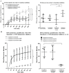
Quantitative analyses show that the NOX-dependent pathway is involved in complement-mediated NET formation. (A) The NET release kinetics were monitored by SYTOX kinetics assay. Complement-stimulated neutrophils release significantly higher amounts of NETs after transitioning from serum to serum-free conditions, compared to other conditions. n = 4 biological replicates; two-way ANOVA. *** p-value < 0.001; * p-value < 0.05. (B) Transferring the complement-activated neutrophils from serum to serum-free condition leads to an increase in ROS generated by NOX but not by mitochondria, as determined by the DHR123 assays. DPI (NOX inhibitor) but not DNP (mitochondrial uncoupler) significantly suppressed ROS generation, as determined by two-way ANOVA with repeated measure, overtime. Comparing the total ROS at a 4 h time point confirmed the differences observed in the kinetics data, as determined by one-way ANOVA with Dunnett’s post-test. * p < 0.05; ** p < 0.01. n = 3 biological replicates. Data are presented as means ± SD.

References

    1. Dunkelberger J.R., Song W.C. Complement and its role in innate and adaptive immune responses. Cell Res. 2010;20:34–50. doi: 10.1038/cr.2009.139. - DOI - PubMed
    1. Neubert E., Senger-Sander S.N., Manzke V.S., Busse J., Polo E., Scheidmann S.E.F., Schön M.P., Kruss S., Serum L.E., Albumin S. Serum and Serum Albumin Inhibit in vitro Formation of Neutrophil Extracellular Traps (NETs) Front. Immunol. 2019;10:12. doi: 10.3389/fimmu.2019.00012. - DOI - PMC - PubMed
    1. Merle N.S., Church S.E., Fremeaux-Bacchi V., Roumenina L.T. Complement System Part I—Molecular Mechanisms of Activation and Regulation. Front. Immunol. 2015;6:262. doi: 10.3389/fimmu.2015.00262. - DOI - PMC - PubMed
    1. Vestweber D. How leukocytes cross the vascular endothelium. Nat. Rev. Immunol. 2015;15:692–704. doi: 10.1038/nri3908. - DOI - PubMed
    1. Noone D.G., Riedl M., Pluthero F.G., Bowman M.L., Liszewski M.K., Lu L., Quan Y., Balgobin S., Schneppenheim R., Schneppenheim S., et al. Von Willebrand factor regulates complement on endothelial cells. Kidney Int. 2016;90:123–134. doi: 10.1016/j.kint.2016.03.023. - DOI - PMC - PubMed

MeSH terms

LinkOut - more resources