. 2024 Sep 7;25(17):9702.
doi: 10.3390/ijms25179702.

Anti-Inflammatory and Anti-(Lymph)angiogenic Properties of an ABCB5+ Limbal Mesenchymal Stem Cell Population

Affiliations

Anti-Inflammatory and Anti-(Lymph)angiogenic Properties of an ABCB5+ Limbal Mesenchymal Stem Cell Population

Berbang Meshko et al. Int J Mol Sci. .

Abstract

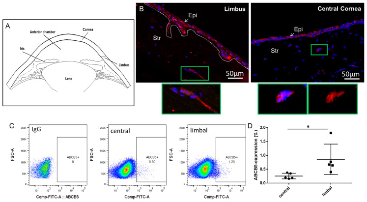
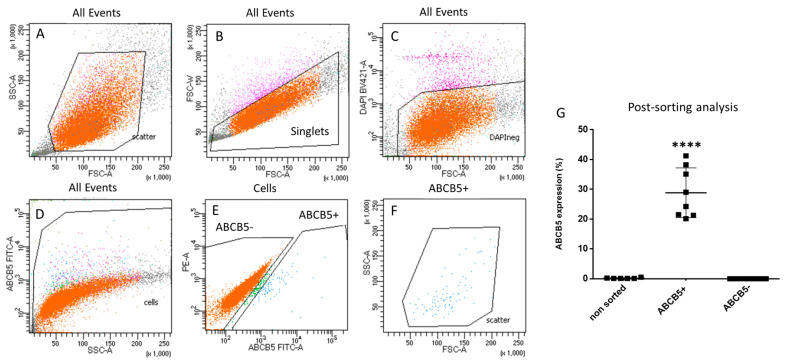
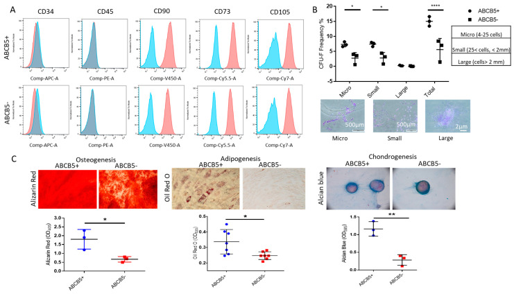
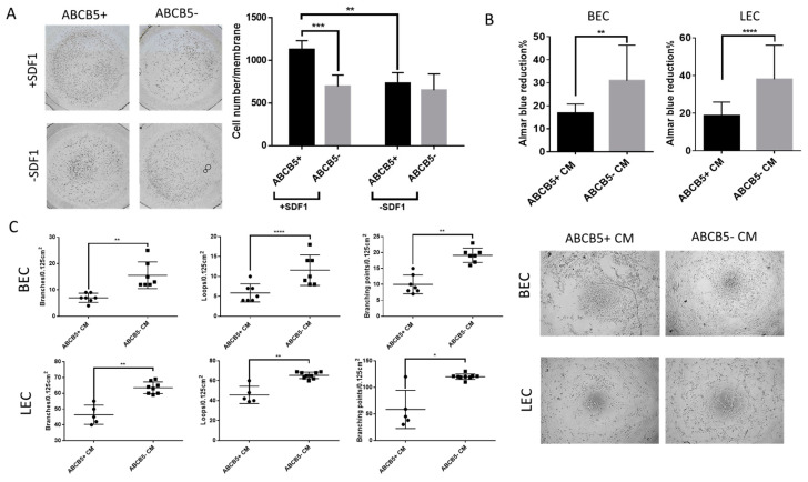
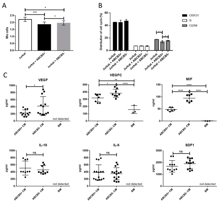
Corneal transparency and avascularity are essential for vision. The avascular cornea transitions into the vascularized conjunctiva at the limbus. Here, we explore a limbal stromal cell sub-population that expresses ABCB5 and has mesenchymal stem cell characteristics. Human primary corneal stromal cells were enriched for ABCB5 by using FACS sorting. ABCB5+ cells expressed the MSC markers CD90, CD73, and CD105. ABCB5+ but not ABCB5- cells from the same donor displayed evidence of pluripotency with a significantly higher colony-forming efficiency and the ability of trilineage differentiation (osteogenic, adipogenic, and chondrogenic). The ABCB5+ cell secretome demonstrated lower levels of the pro-inflammatory protein MIF (macrophage migration inhibitory factor) as well as of the pro-(lymph)angiogenic growth factors VEGFA and VEGFC, which correlated with reduced proliferation of Jurkat cells co-cultured with ABCB5+ cells and decreased proliferation of blood and lymphatic endothelial cells cultured in ABCB5+ cell-conditioned media. These data support the hypothesis that ABCB5+ limbal stromal cells are a putative MSC population with potential anti-inflammatory and anti-(lymph)angiogenic effects. The therapeutic modulation of ABCB5+ limbal stromal cells may prevent cornea neovascularization and inflammation and, if transplanted to other sites in the body, provide similar protective properties to other tissues.

Keywords: (lymph)angiogenesis; ABCB5; cornea; mesenchymal stem cells.


Conflict of interest statement

C.G.: CEO of RHEACELL GmbH & Co. KG. M.A.K.: CSO of RHEACELL GmbH & Co. KG. M.H.F., B.R.K., and N.Y.F. are inventors or co-inventors of US and international patents assigned to Brigham and Women’s Hospital, Boston Children’s Hospital, the Massachusetts Eye and Ear Infirmary, and/or the VA Boston Healthcare System, Boston, MA, licensed to RHEACELL GmbH & Co. KG (Heidelberg, Germany). M.H.F. serves as a scientific advisor to and holds stock in RHEACELL GmbH & Co. KG.

Figures

Figure 1
Figure 1
ABCB5 is expressed in limbal and central corneal stromal cells. (A) A schematic of the cornea cross section depicting the locations of the limbus, cornea, lens, and iris. (B) Immunocytochemistry of limbal and central corneal sections, red: ABCB5 (alexa 555) blue: DAPI. In green frame: magnified insets depicting clusters of ABCB5-expressing stromal cells in the limbal stroma and cornea stroma, respectively. Images captured using 20x magnification. (C) ABCB5 expression evaluated by FACS analysis of expanded stromal cells from the central cornea and the limbus. (D) Quantification of FACS data comparing the percentage of ABCB5-expresing cells derived from the central cornea and the limbus (* signifies p < 0.05, n = 5).
Figure 2
Figure 2
FACS sorting gating strategy for selection of ABCB5+ cells. (A) Cells separation from debris (forward scatter-FSC/side scatter-SSC dot-plot). (B) Exclusion of doublets. (C) Selection of live cells and (D,E) definition of ABCB5+ and ABCB5- cells. (F) Back-gating of the ABCB5+ gate to the SSC and FSC plot. (G) Post-sorting FACS analysis results (**** signifies p < 0.0001, n = 8).
Figure 3
Figure 3
Comparison of MSC properties of ABCB5+ and ABCB5−cells. (A) Representative histograms of FACS analysis for a mesenchymal stem cell (positive and negative) marker panel including CD34, CD45, CD90, CD73, and CD105, demonstrating similar expression profiles for both ABCB5+ and ABCB5− cells. Blue: IgG control, Red: stained samples (B) Colony-forming unit efficiency comparing the percentage of micro-, small, large, and total colonies generated by ABCB5+ or ABCB5- cells and representative colony photos. (C) Trilineage differentiation assays using Alizarin Red (Osteogenesis), Oil Red O (Adipogenesis), and Alcian Blue (Chondrogenesis), and respective quantification of extracted dye solutions by absorbance measurements (n ≥ 3). Images captured using 20x magnification. In all graphs: * signifies p < 0.05, ** signifies p < 0.01 and **** signifies p < 0.0001.
Figure 4
Figure 4
ABCB5+ cells exhibit increased migratory activity while they hamper proliferation and tube formation of blood and lymphatic endothelial cells. (A) Representative photographs of migrated ABCB5+ and ABCB5- cells in the presence or absence of SDF1 and respective graph depicting quantification of the migrated cell numbers (n = 5). Images captured using 5x magnification. (B) Alamar Blue (metabolic activity) assay of blood and lymphatic endothelial cells (BEC and LEC) treated with conditioned media from ABCB5+ and ABCB5- cells (n = 7). (C) Tube-formation assay representative photos and morphometric analysis by evaluation of branches, loops, and branching points of BECs and LECs treated with conditioned media from ABCB5+ and ABCB5- cells (ABCB5+ CM and ABCB5- CM, n = 3). Images captured using 20x magnification. In all graphs: * signifies p < 0.05, ** signifies p < 0.01, *** signifies p < 0.001 and **** signifies p < 0.0001.
Figure 5
Figure 5
Co-culture of ABCB5+ and ABCB5- cells with Jurkat cells and ELISA of proangiogenic and proinflammatory cytokines demonstrate a comparative anti-inflammatory effect and anti-inflammatory and anti-angiogenic secreted cytokine profile of ABCB5+ cells. (A) Metabolic activity (Alamar Blue assay) and (B) cell cycle analysis of Jurkat cells co-cultured with ABCB5+ and ABCB5- cells (24 h). (C) Evaluation by ELISA of VEGFA, VEGFC, MIF, IL-18, IL-6, and SDF1 secreted in conditioned media of ABCB5+ and ABCB5- cells (ABCB5+ CM and ABCB5- CM, n = 5). * signifies p < 0.05, ** signifies p < 0.01, *** signifies p < 0.001 and **** signifies p < 0.0001.

References

    1. Notara M., Alatza A., Gilfillan J., Harris A.R., Levis H.J., Schrader S., Vernon A., Daniels J.T. In sickness and in health: Corneal epithelial stem cell biology, pathology and therapy. Exp. Eye Res. 2010;90:188–195. doi: 10.1016/j.exer.2009.09.023. - DOI - PubMed
    1. DelMonte D.W., Kim T. Anatomy and physiology of the cornea. J. Cataract Refract. Surg. 2011;37:588–598. doi: 10.1016/j.jcrs.2010.12.037. - DOI - PubMed
    1. Feuk T. On the transparency of the stroma in the mammalian cornea. IEEE Trans. Bio-Med. Eng. 1970;17:186–190. doi: 10.1109/TBME.1970.4502732. - DOI - PubMed
    1. Hanlon S.D., Behzad A.R., Sakai L.Y., Burns A.R. Corneal stroma microfibrils. Exp. Eye Res. 2015;132:198–207. doi: 10.1016/j.exer.2015.01.014. - DOI - PMC - PubMed
    1. Meek K.M., Knupp C., Lewis P.N., Morgan S.R., Hayes S. Structural control of corneal transparency, refractive power and dynamics. Eye. 2024;1 doi: 10.1038/s41433-024-02969-7. - DOI - PMC - PubMed

MeSH terms

Substances

LinkOut - more resources