. 2024 Aug 30;16(17):2470.
doi: 10.3390/polym16172470.

Coaxial Bioprinting of Enzymatically Crosslinkable Hyaluronic Acid-Tyramine Bioinks for Tissue Regeneration

Affiliations

Coaxial Bioprinting of Enzymatically Crosslinkable Hyaluronic Acid-Tyramine Bioinks for Tissue Regeneration

Alma Tamunonengiofori Banigo et al. Polymers (Basel). .

Abstract

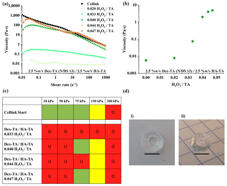
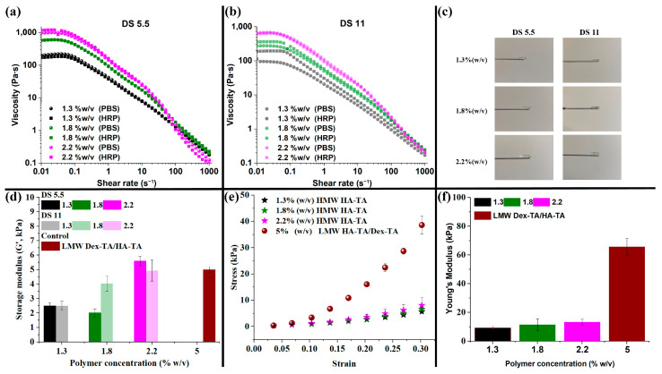
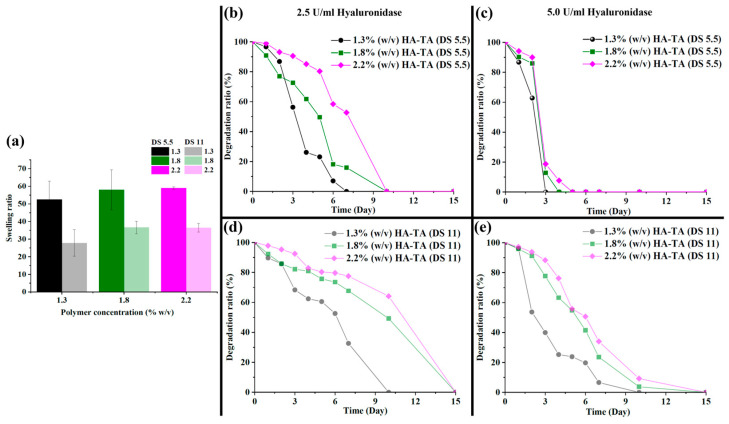
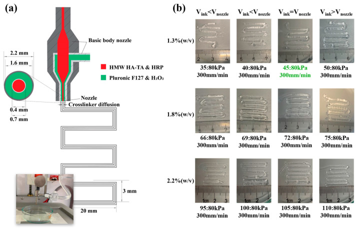
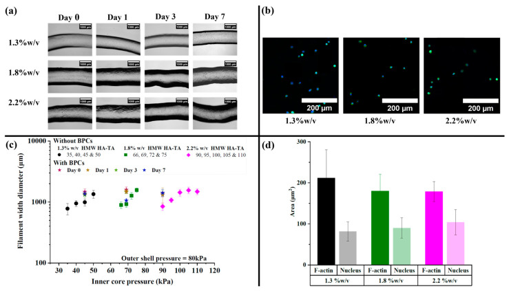
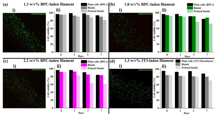
Three-dimensional (3D) bioprinting has emerged as an important technique for fabricating tissue constructs with precise structural and compositional control. However, developing suitable bioinks with biocompatible crosslinking mechanisms remains a significant challenge. This study investigates extrusion-based bioprinting (EBB) using uniaxial or coaxial nozzles with enzymatic crosslinking (EC) to produce 3D tissue constructs in vitro. Initially, low-molecular-weight dextran-tyramine and hyaluronic acid-tyramine (LMW Dex-TA/HA-TA) bioink prepolymers were evaluated. Enzymatically pre-crosslinking these prepolymers, achieved by the addition of horseradish peroxidase and hydrogen peroxide, produced viscous polymer solutions. However, this approach resulted in inconsistent bioprinting outcomes (uniaxial) due to inhomogeneous crosslinking, leading to irreproducible properties and suboptimal shear recovery behavior of the hydrogel inks. To address these challenges, we explored a one-step coaxial bioprinting system consisting of enzymatically crosslinkable high-molecular-weight hyaluronic acid-tyramine conjugates (HMW HA-TA) mixed with horseradish peroxidase (HRP) in the inner core and a mixture of Pluronic F127 and hydrogen peroxide in the outer shell. This configuration resulted in nearly instantaneous gelation by diffusion of the hydrogen peroxide into the core. Stable hydrogel fibers with desirable properties, including appropriate swelling ratios and controlled degradation rates, were obtained. The optimized bioink and printing parameters included 1.3% w/v HMW HA-TA and 5.5 U/mL HRP (bioink, inner core), and 27.5% w/v Pluronic F127 and 0.1% H2O2 (sacrificial ink, outer shell). Additionally, optimal pressures for the inner core and outer shell were 45 and 80 kPa, combined with a printing speed of 300 mm/min and a bed temperature of 30 °C. The extruded HMW HA-TA core filaments, containing bovine primary chondrocytes (BPCs) or 3T3 fibroblasts (3T3 Fs), exhibited good cell viabilities and were successfully cultured for up to seven days. This study serves as a proof-of-concept for the one-step generation of core filaments using a rapidly gelling bioink with an enzymatic crosslinking mechanism, and a coaxial bioprinter nozzle system. The results demonstrate significant potential for developing designed, printed, and organized 3D tissue fiber constructs.

Keywords: bioink; coaxial bioprinting; enzymatic crosslinking; hyaluronic acid-tyramine conjugates; tissue regeneration.


Conflict of interest statement

The authors declare no conflict of interest.

Figures

Figure 1
Figure 1
Rheological analysis and printing conditions of 5% w/v LMW Dex-TA/HA-TA (Dex-TA: N/DS 13) with 1 U/mL HRP. (a) Viscosity during a shear rate sweep from 0.01 s−1 to 1000 s−1 of LMW Dex-TA/HA-TA solutions pre-crosslinked at different H2O2/TA ratios, represented by the numbers in the legend. (b) Viscosity at 100 s−1 shear as a function of the H2O2/TA ratio. As shown in the plot, viscosity was positively correlated with an increase in the H2O2/TA ratio. At higher H2O2/TA ratios of >0.04, the viscosity increase seemed to reach a plateau. (c) Printing studies of bioinks at different printing pressures and a single nozzle of 25 G. The printing quality is identified with distinct colors: good (green, <200% filament spread and <10% diameter variation), moderate (yellow, 200–300% filament spread and >10% diameter variation), or poor (red, divided into under-extrusion (U) and over-extrusion (O)) printability, depending on the criteria, as shown in the legend. (d) The 3D-printed hollow cylinder (i) top view and (ii) side view. The grid lines in image (ii) comprise of 5 mm × 5 mm (scale bars represent 5000 µm).
Figure 2
Figure 2
Shear-thinning and mechanical properties of HMW HA-TA bioinks and corresponding hydrogels. Shear-thinning profile of HMW HA-TA with (a) DS 5.5 and (b) DS 11 at polymer concentrations of 1.3, 1.8, and 2.2% w/v. (c) Hydrogels produced with 5.5 U/mL HRP and a 0.5 H2O2:TA molar ratio. (d) Storage modulus of all hydrogel compositions, including a 5% w/v LMW Dex-TA/HA-TA control hydrogel. (e) Stress–strain curves of DS 5.5 hydrogels compared with a 5% w/v LMW Dex-TA/HA-TA control hydrogel. (f) Young’s modulus of the cylindrical hydrogels.
Figure 3
Figure 3
Physical properties of the HMW HA-TA hydrogels. (a) Swelling ratio of the HMW HA-TA hydrogels. (b) Degradation of the HMW HA-TA hydrogels with (b) DS 5.5 in 2.5 U/mL hyaluronidase, (c) DS 5.5 in 5 U/mL hyaluronidase, (d) DS 11 in 2.5 U/mL hyaluronidase, and (e) DS 11 in 5 U/mL hyaluronidase.
Figure 4
Figure 4
(a) Schematic representation of the coaxial 3D-printing system with a cross-sectional view of the coaxial nozzle tips and programmed pattern designed for printing. (b) Coaxially printed core filaments of 1.3, 1.8, and 2.2% w/v HMW HA-TA with different inner core pressures, a fixed outer shell pressure (80 kPa), and the same printing speed (300 mm/min); conditions for a speed match are indicated in green.
Figure 5
Figure 5
Core filaments with cells. (a) BPC-laden core filaments printed with 1.3, 1.8, and 2.2% w/v HMW HA-TA bioinks containing 5.5 U/mL HRP, printed at 45, 69, and 90 kPa, respectively (scale bars represent 1000 µm). (b) The BPCs were stained for F-actin and nuclei on day 7, revealing their rounded shape (scale bars represent 200 µm). (c) Quantified filament width of the core filaments with and without BPCs. (d) Quantified area of the rounded F-actin and nuclei.
Figure 6
Figure 6
Core filaments with cells. (aici) Confocal fluorescence images of printed BPC-laden filaments of 1.3, 1.8, and 2.2% w/v HMW HA-TA, respectively. (di) Confocal fluorescence images of printed 3T3 fibroblast-laden filaments of 1.3% w/v HA-TA (scale bars represents 800 µm). (aiidii) Corresponding cell viability plots of plain cells (BPCs and 3T3 fibroblasts), extruded bioinks, and printed bioinks (crosslinked filaments).

Similar articles

Cited by

References

    1. Dai X., Liu L., Ouyang J., Li X., Zhang X., Lan Q., Xu T. Coaxial 3D bioprinting of self-assembled multicellular heterogeneous tumor fibers. Sci. Rep. 2017;7:1457. doi: 10.1038/s41598-017-01581-y. - DOI - PMC - PubMed
    1. Gao G., Kim B.S., Jang J., Cho D.-W. Recent Strategies in Extrusion-Based Three-Dimensional Cell Printing toward Organ Biofabrication. ACS Biomater. Sci. Eng. 2019;5:1150–1169. doi: 10.1021/acsbiomaterials.8b00691. - DOI - PubMed
    1. Pati F., Jang J., Ha D.-H., Kim S.W., Rhiie J.-W., Shim J.-H., Kim D.-H., Cho D.-W. Printing three-dimensional tissue analogues with decellularized extracellular matrix bioink. Nat. Commun. 2014;5:3935. doi: 10.1038/ncomms4935. - DOI - PMC - PubMed
    1. Mironov V., Trusk T., Kasyanov V., Little S., Swaja R., Markwald R. Biofabrication: A 21st century manufacturing paradigm. Biofabrication. 2009;1:022001. doi: 10.1088/1758-5082/1/2/022001. - DOI - PubMed
    1. Petta D., Armiento A.R., Grijpma D., Alini M., Eglin D., D’este M. 3D bioprinting of a Hyaluronan bioink through enzymatic-and visible light-crosslinking. Biofabrication. 2018;10:044104. doi: 10.1088/1758-5090/aadf58. - DOI - PubMed

LinkOut - more resources