

Development and Validation of Artificial Intelligence Models for Prognosis Prediction of Juvenile Myoclonic Epilepsy with Clinical and Radiological Features
- PMID: 39274294
- PMCID: PMC11396353
- DOI: 10.3390/jcm13175080
Development and Validation of Artificial Intelligence Models for Prognosis Prediction of Juvenile Myoclonic Epilepsy with Clinical and Radiological Features
Abstract
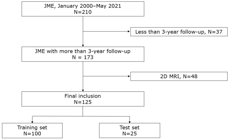
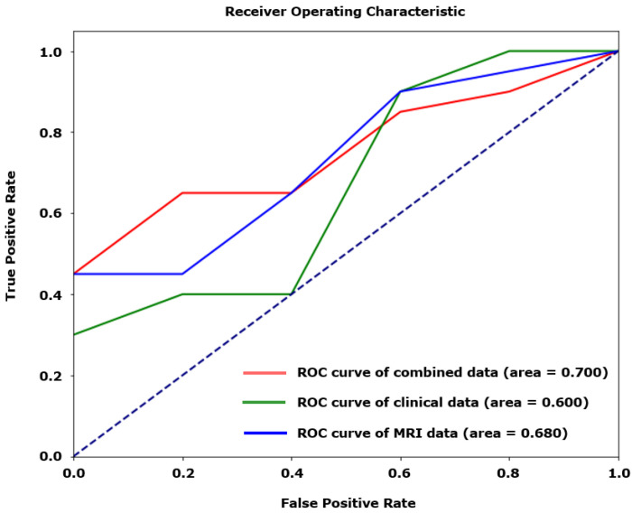
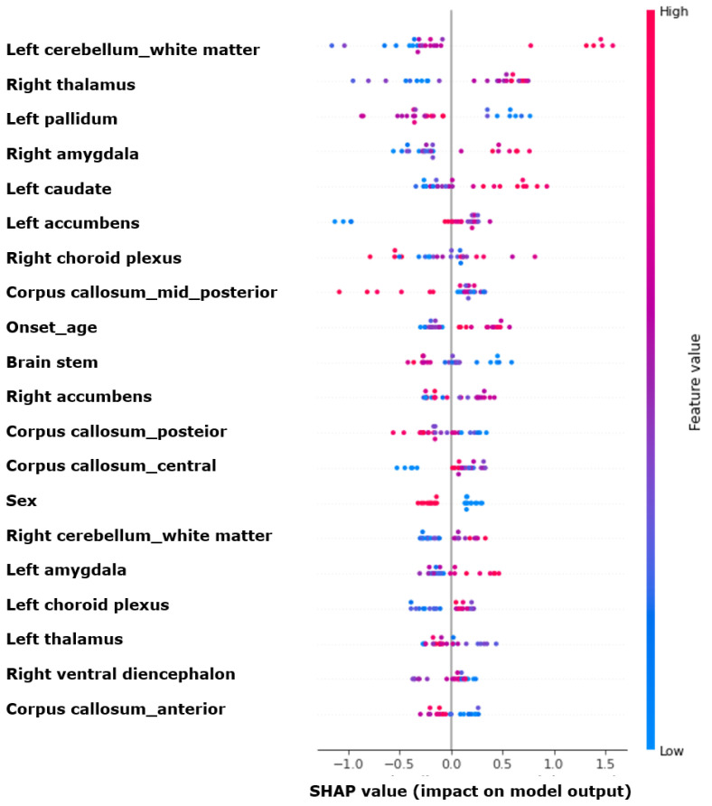
Background: Juvenile myoclonic epilepsy (JME) is a common adolescent epilepsy characterized by myoclonic, generalized tonic-clonic, and sometimes absence seizures. Prognosis varies, with many patients experiencing relapse despite pharmacological treatment. Recent advances in imaging and artificial intelligence suggest that combining microstructural brain changes with traditional clinical variables can enhance potential prognostic biomarkers identification. Methods: A retrospective study was conducted on patients with JME at the Severance Hospital, analyzing clinical variables and magnetic resonance imaging (MRI) data. Machine learning models were developed to predict prognosis using clinical and radiological features. Results: The study utilized six machine learning models, with the XGBoost model demonstrating the highest predictive accuracy (AUROC 0.700). Combining clinical and MRI data outperformed models using either type of data alone. The key features identified through a Shapley additive explanation analysis included the volumes of the left cerebellum white matter, right thalamus, and left globus pallidus. Conclusions: This study demonstrated that integrating clinical and radiological data enhances the predictive accuracy of JME prognosis. Combining these neuroanatomical features with clinical variables provided a robust prediction of JME prognosis, highlighting the importance of integrating multimodal data for accurate prognosis.
Keywords: artificial intelligence; juvenile myoclonic epilepsy; machine learning; magnetic resonance imaging; prognosis.
Conflict of interest statement
All the authors have no conflicts to disclose.
Figures
Similar articles
-
MRI-Based Radiomics Approach for Differentiating Juvenile Myoclonic Epilepsy from Epilepsy with Generalized Tonic-Clonic Seizures Alone.J Magn Reson Imaging. 2024 Jul;60(1):281-288. doi: 10.1002/jmri.29024. Epub 2023 Oct 10. J Magn Reson Imaging. 2024. PMID: 37814782
-
Subcortical gray matter changes in pediatric patients with new-onset juvenile myoclonic epilepsy.Epilepsy Behav. 2020 Mar;104(Pt A):106860. doi: 10.1016/j.yebeh.2019.106860. Epub 2020 Jan 11. Epilepsy Behav. 2020. PMID: 31935646
-
Subcortical gray matter changes in juvenile myoclonic epilepsy: a volBrain study.Folia Morphol (Warsz). 2025 Mar 27. doi: 10.5603/fm.103357. Online ahead of print. Folia Morphol (Warsz). 2025. PMID: 40145711
-
Gray Matter Changes in Juvenile Myoclonic Epilepsy. A Voxel-Wise Meta-Analysis.Medicina (Kaunas). 2021 Oct 20;57(11):1136. doi: 10.3390/medicina57111136. Medicina (Kaunas). 2021. PMID: 34833354 Free PMC article. Review.
-
Grey and White Matter Alterations in Juvenile Myoclonic Epilepsy: A Comprehensive Review.J Epilepsy Res. 2017 Dec 31;7(2):77-88. doi: 10.14581/jer.17013. eCollection 2017 Dec. J Epilepsy Res. 2017. PMID: 29344465 Free PMC article. Review.
References
-
- Proposal for classification of epilepsies and epileptic syndromes Commission on classification and terminology of the international league against epilepsy. Epilepsia. 1985;26:268–278. - PubMed
Grants and funding
LinkOut - more resources
Full Text Sources
