

Cone-Beam Computed Tomography (CBCT) Analysis of Mandibular Condyles' Diameters in Patient with Juvenile Idiopathic Arthritis and Temporomandibular Joint Affection: A Cross-Sectional Investigation
- PMID: 39274322
- PMCID: PMC11395992
- DOI: 10.3390/jcm13175104
Cone-Beam Computed Tomography (CBCT) Analysis of Mandibular Condyles' Diameters in Patient with Juvenile Idiopathic Arthritis and Temporomandibular Joint Affection: A Cross-Sectional Investigation
Abstract
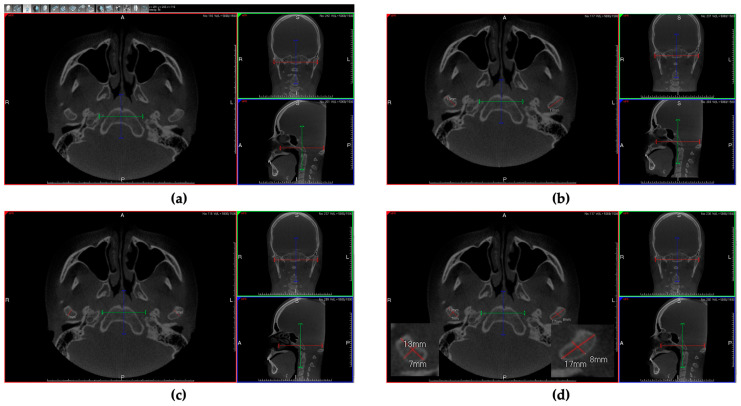
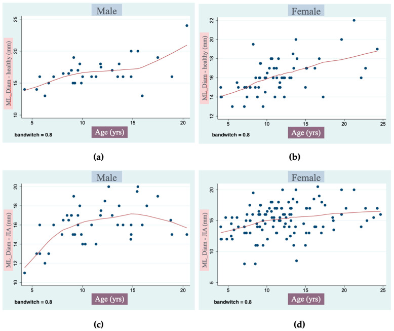
Objectives: The aim of this study was to evaluate through analysis using CBCT the transverse and axial diameters of the mandibular condyles in subjects affected by juvenile idiopathic arthritis (JIA) and compare them with those of healthy subjects. Methods: The study was conducted on CBCT scans from the digital archive of the Department of Biomedical Surgical and Dental Sciences, University of Milan, including patients with JIA and using healthy subjects as controls. Inclusion criteria: aged between 7 and 25 years old at the time of the CBCT examination; Caucasian ethnicity; diagnosis of JIA according to the International League of Associations for Rheumatology (ILAR) criteria documented in patients' records; TMJ involvement; good quality CBCTs covering our region of interest (ROI), from the glabella to the mandibular inferior border; no previous orthodontic/orthopedic treatment; no history of craniofacial trauma or congenital birth defects involving the craniofacial area. Each CBCT scan underwent examination using 3Diagnosys® software. Since data were normally distributed, parametric tests were used for analysis. The sample was divided into three groups: (1) bilateral JIA subjects, (2) unilateral JIA subjects, and (3) healthy controls. Results: We found a statistically significant reduction (p < 0.0001) in the transverse diameter (TR-Diam) of the affected condyles by an average of 1.7 mm, while the axial diameter (AX-Diam) again showed a slight reduction, on average by 0.1 mm, with a non-statistically significant value. Another comparison was made between the unaffected condyles of patients with unilateral JIA and the healthy condyles of the control group. The unilateral unaffected condyles were found to be slightly smaller than those of healthy patients, but without statistically significant differences. We found that in both JIA males and females, the condylar growth tends to stop earlier than the healthy ones. Conclusions: The transverse diameter was found to be more affected than the axial one, causing typical bone resorption and condylar shape. Moreover, we showed that the pathology, in the case of unilateral JIA, does not compromise only the affected condyles; the corresponding condyle that seems to be healthy is actually partially compromised. In addition, we observed that the growth of affected condyles of JIA subjects tends to stop earlier than the condyles of the healthy controls.
Keywords: CBCT; juvenile idiopathic arthritis; malocclusion; mandibular condyle; temporomandibular joint arthritis.
Conflict of interest statement
The authors declare no conflicts of interest.
Figures
Similar articles
-
Assessment of condylar volume and ramus height in JIA patients with unilateral and bilateral TMJ involvement: retrospective case-control study.Clin Oral Investig. 2020 Aug;24(8):2635-2643. doi: 10.1007/s00784-019-03122-5. Epub 2019 Nov 23. Clin Oral Investig. 2020. PMID: 31760476
-
Three-dimensional volumetric evaluation of the different mandibular segments using CBCT in patients affected by juvenile idiopathic arthritis: a cross-sectional study.Prog Orthod. 2021 Oct 1;22(1):32. doi: 10.1186/s40510-021-00380-6. Prog Orthod. 2021. PMID: 34595615 Free PMC article.
-
Comparison of the condyle-fossa relationship and resorption between patients with and without Juvenile Idiopathic Arthritis (JIA).J Oral Maxillofac Surg. 2022 Mar;80(3):422-430. doi: 10.1016/j.joms.2021.09.004. Epub 2021 Sep 12. J Oral Maxillofac Surg. 2022. PMID: 34627744
-
What is the image appearance of juvenile idiopathic arthritis in MRI, CT, and CBCT of TMJ? A systematic review.Clin Oral Investig. 2023 May;27(5):2321-2333. doi: 10.1007/s00784-022-04828-9. Epub 2022 Dec 14. Clin Oral Investig. 2023. PMID: 36515761
-
Management of temporomandibular joint arthritis in children and adolescents: An introduction for orthodontists.Orthod Craniofac Res. 2023 Dec;26 Suppl 1:151-163. doi: 10.1111/ocr.12676. Epub 2023 May 24. Orthod Craniofac Res. 2023. PMID: 37226648 Review.
Cited by
-
Changes of Airway Space and Flow in Patients Treated with Rapid Palatal Expander (RPE): An Observational Pilot Study with Comparison with Non-Treated Patients.J Clin Med. 2025 Jun 18;14(12):4357. doi: 10.3390/jcm14124357. J Clin Med. 2025. PMID: 40566102 Free PMC article.
References
-
- Petty R.E., Southwood T.R., Manners P., Baum J., Glass D.N., Goldenberg J., He X., Maldonado-Cocco J., Orozco-Alcala J., Prieur A.-M., et al. International League of Associations for Rheumatology Classification of Juvenile Idiopathic Arthritis: Second Revision, Edmonton, 2001. J. Rheumatol. 2004;31:390–392. - PubMed
-
- Stoustrup P., Resnick C.M., Pedersen T.K., Abramowicz S., Michelotti A., Küseler A., Verna C., Kellenberger C.J., Berit Nordal E., Caserta G., et al. Standardizing Terminology and Assessment for Orofacial Conditions in Juvenile Idiopathic Arthritis: International, Multidisciplinary Consensus-Based Recommendations. J. Rheumatol. 2019;46:518–522. doi: 10.3899/jrheum.180785. - DOI - PubMed
-
- Farronato M., Cavagnetto D., Abate A., Cressoni P., Fama A., Maspero C. Assessment of Condylar Volume and Ramus Height in JIA Patients with Unilateral and Bilateral TMJ Involvement: Retrospective Case-Control Study. Clin. Oral Investig. 2020;24:2635–2643. doi: 10.1007/s00784-019-03122-5. - DOI - PubMed
LinkOut - more resources
Full Text Sources
