

Population Analysis of Masseter Muscle Tension Using Shear Wave Ultrasonography across Different Disease States
- PMID: 39274477
- PMCID: PMC11396082
- DOI: 10.3390/jcm13175259
Population Analysis of Masseter Muscle Tension Using Shear Wave Ultrasonography across Different Disease States
Abstract
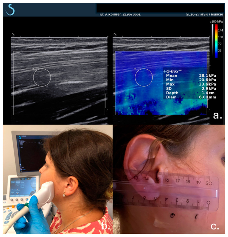
Objective: This study aimed to evaluate the distribution and trends of masseter muscle tension in patients with temporomandibular joint (TMJ) pain, examining gender-specific differences and the impact of various TMJ disorders. Methods: From January 2020 to June 2024, a total of 734 patients presenting with facial pain radiating to the head and neck, localized around and extending from the TMJ, were referred for ultrasonographic examination. After applying exclusion criteria, 535 patients (72.9%) were included in the study. The patient cohort consisted of 343 females (64.1%) and 192 males (35.9%), with muscle tension measured using the Aixplorer ultrasound system equipped with a shear wave device. Data were collected and analyzed across different age groups and TMJ conditions, including "no changes", "exudate", "arthrosis", and "disc displacement". Results: The study found that males exhibited higher muscle tension across all conditions, particularly in the "no changes" (40.4 kPa vs. 32.1 kPa, 25.9% higher) and "exudate" (38.5 kPa vs. 29.7 kPa, 29.6% higher) categories, indicating increased muscle strain and inflammation during middle age. In females, a trend of decreasing muscle tension with age was observed, with a significant reduction from 36.2 kPa in the 20-30 age group to 24.3 kPa in the 60-70 age group (32.9% reduction), suggesting a reduction in muscle mass or strength due to aging. Both genders showed high muscle tension in the presence of exudate, with females peaking in the 40-50 age group at 37.1 kPa and males peaking earlier in the 20-30 age group at 41.2 kPa (10.9% higher in males), highlighting potential gender differences in inflammatory response. In the arthrosis group, males displayed a consistent increase in muscle tension with age, peaking at 37.5 kPa in the 50-60 age group (50.7% increase from the 20-30 age group), while females showed high tension, particularly in the 40-50 age group at 31.0 kPa (82.4% higher compared to the 20-30 age group), indicating the need for targeted joint health interventions in middle-aged women. Conclusions: This study reveals significant gender-specific differences in masseter muscle tension among patients with TMJ pain. Males were found to be more affected by muscle strain and inflammation during middle age, whereas females showed a significant decrease in muscle tension with age. The presence of exudate significantly impacted muscle tension across all age groups for both genders. These findings underscore the importance of tailored clinical interventions and preventive strategies to manage TMJ disorders effectively.
Keywords: elastography; masseter muscle; shear wave; temporomandibular disorders.
Conflict of interest statement
The authors declare no conflicts of interest.
Figures


References
-
- Sarnat B.G., Laskin D.M. The Temporomandibular Joint: A Biological Basis for Clinical Practice. 4th ed. WB Saunders; Philadelphia, PA, USA: 1992. pp. 78–163.
-
- Graber R., Rakosi T. Dentofacial Orthopedics with Functional Appliances. 2nd ed. Mosby; St. Louis, MO, USA: 2009. Petrovic. Functional analysis- examination of temporomandibular joint and condylar movement; pp. 135–140.
-
- Athanasiou A.E. Orthodontics and craniomandibular disorders. In: Samire B., editor. Textbook of Orthodontics. 2nd ed. Saunders; Philadelphia, PA, USA: 2003. pp. 478–493.
-
- Irby W.B., Baldwin K.H. Emergencies and Urgent Complications in Dentistry. The C. V. Mosby Company; St. Louis, MO, USA: 1965. pp. 167–173.
-
- Donovan T.E., Becker W., Brodine A.H., Burgess J.O., Cronin R.J., Summitt J.B. Annual review of selected dental literature: Report of the Committee on Scientific Investigation of the American Academy of Restorative Dentistry. J. Prosthet. Dent. 2007;98:36–67. doi: 10.1016/S0022-3913(07)60036-5. - DOI - PubMed
LinkOut - more resources
Full Text Sources
