. 2024 Aug 24;24(17):5481.
doi: 10.3390/s24175481.

SPRi Biosensor for Simultaneous Determination of HIF-1α, Angiopoietin-2, and Interleukin-1β in Blood Plasma

Affiliations

SPRi Biosensor for Simultaneous Determination of HIF-1α, Angiopoietin-2, and Interleukin-1β in Blood Plasma

Zuzanna Zielinska et al. Sensors (Basel). .

Abstract

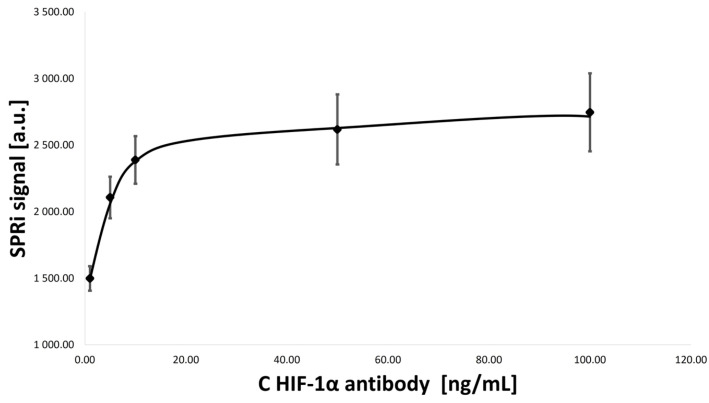
A new analytical method, based on SPRi biosensors, has been developed for the simultaneous determination of the pro-angiogenic factors HIF-1α, angiopoietin-2 (ANG-2), and interleukin-1β (IL-1β) in biological fluids. These proteins take part in the process of angiogenesis, i.e., the creation of new blood vessels, which is a key stage of cancer development and metastasis. A separate validation process was carried out for each individual compound, indicating that the method can also be used to study one selected protein. Low values of the limit of detection (LOD) and quantification (LOQ) indicate that the developed method enables the determination of very low concentrations, in the order of pg/mL. The LOD values obtained for HIF-1α, ANG-2, and IL-1β were 0.09, 0.01, and 0.01 pg/mL, respectively. The LOQ values were 0.27, 0.039, and 0.02 pg/mL, and the response ranges of the biosensor were 5.00-100.00, 1.00-20.00, and 1.00-15.00 pg/mL. Moreover, determining the appropriate validation parameters confirmed that the design offers high precision, accuracy, and sensitivity. To prove the usefulness of the biosensor in practice, determinations were made in plasma samples from a control group and from a study group consisting of patients with diagnosed bladder cancer. The preliminary results obtained indicate that this biosensor can be used for broader analyses of bladder cancer. Each of the potential biomarkers, HIF-1α, ANG-2, and IL-1β, produced higher concentrations in the study group than in the control group. These are preliminary studies that serve to develop hypotheses, and their confirmation requires the analysis of a larger number of samples. However, the constructed biosensor is characterized by its ease and speed of measurement, and the method does not require special preparation of samples. SPRi biosensors can be used as a sensitive and highly selective method for determining potential blood biomarkers, which in the future may become part of the routine diagnosis of cancers.

Keywords: angiogenesis; biosensor; surface plasmon resonance.


Conflict of interest statement

The authors declare no conflicts of interest.

Figures

Figure A1
Figure A1
Steps for immobilization of the antibody layer (illustration by the authors). To immobilize the antibody on the gold surface with a thiol layer, a mixture of EDC and NHS is applied in a 1:1 volume ratio. The carboxyl group of the thiol is converted into an ester group by EDC, while NHS modifies the ester into a short-lived NHS-ester. In the next step, the applied antibody forms a covalent amide bond with the NHS-ester. To prevent non-specific adsorption, ethanolamine is applied, which deactivates active NHS-esters and also prevents the attachment of other components to thiol molecules that have not bound to the antibody.
Figure A2
Figure A2
Antibody saturation curve for HIF-1α.
Figure A3
Figure A3
Antibody saturation curve for ANG-2.
Figure A4
Figure A4
Antibody saturation curve for IL-1β.
Figure 1
Figure 1
HIF-1α protein structure [9].
Figure 2
Figure 2
ANG-2 protein structure [18].
Figure 3
Figure 3
IL-1β protein structure [33].
Figure 4
Figure 4
Schematic diagram of the SPRi apparatus with device components. The LED laser emits a beam of light that passes through a system of polarizers and lenses and then hits a prism with a biosensor, a plate with a gold layer. During measurement, the light is collected by a CCD camera and sent to a computer. Own elaboration.
Figure 5
Figure 5
Construction of the biosensor for the determination of HIF-1α, ANG-2, and IL-1β (A—BK7 glass layer; B—titanium layer; C—gold layer; D—polymer foil; E—thiol–mercaptoundecanoic acid 11-MUA; F1—IL-1β-specific antibody; F2—ANG-2-specific antibody; F3—HIF-1α-specific antibody; G1—IL-1β protein; G2—ANG-2 protein; G3—HIF-1α protein). Each generated receptor layer (thiol–antibody) is separate for a given protein. In this way, three measurement spots for detecting each protein were created on one chip, which can be determined simultaneously in one measurement cycle. Own elaboration.
Figure 6
Figure 6
The course of model SPR curves. For each of the distinguished layers present on the biosensor surface, data were collected at angles from 34 to 37 degrees, in 0.1-degree increments. Each of the curves for a given layer is shifted towards larger angle values relative to the curve of the preceding layer.
Figure 7
Figure 7
HIF-1α, ANG-2, and IL-1β calibration curves. Ligand/antibody concentration for HIF-1α = 50.00 ng/mL, ligand/antibody concentration for ANG-2 = 5.00 μg/mL, and ligand/antibody concentration for IL-1β = 100.00 ng/mL. Calibration curves were prepared using standard solutions prepared in PBS buffer.

References

    1. Lugano R., Ramachandran M., Dimberg A. Tumor angiogenesis: Causes, consequences, challenges and opportunities. Cell. Mol. Life Sci. 2020;77:1745–1770. doi: 10.1007/s00018-019-03351-7. - DOI - PMC - PubMed
    1. Shi Y.-H., Fang W.-G. Hypoxia-inducible factor-1 in tumour angiogenesis. World J. Gastroenterol. 2004;10:1082. doi: 10.3748/wjg.v10.i8.1082. - DOI - PMC - PubMed
    1. Singh R.P., Agarwal R. Tumor Angiogenesis: A Potential Target In Cancer Control by Phytochemicals. Curr. Cancer Drug Targets. 2003;3:205–217. doi: 10.2174/1568009033481985. - DOI - PubMed
    1. Czajka I. Angiogeneza nowotworowa. Endokrynol. Pol. 2004;4:454–455.
    1. Szala S., Markowska J. In: Naczynia Okołonowotworowe jako Cele Terapii Przeciwnowotworowej. Markowska J., editor. Wydawnictwo Medyczne Urban & Partner; Wrocław, Poland: 2006. pp. 44–57. Ginekologia Onkologiczna.

MeSH terms

LinkOut - more resources