. 2024 Sep 4;24(17):5749.
doi: 10.3390/s24175749.

Variational Mode Decomposition Analysis of Electroencephalograms during General Anesthesia: Using the Grey Wolf Optimizer to Determine Hyperparameters

Affiliations

Variational Mode Decomposition Analysis of Electroencephalograms during General Anesthesia: Using the Grey Wolf Optimizer to Determine Hyperparameters

Kosuke Kushimoto et al. Sensors (Basel). .

Abstract

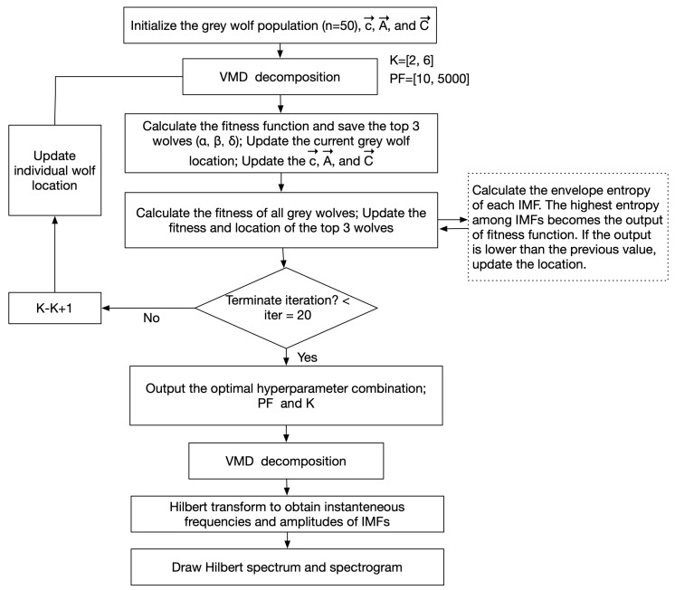
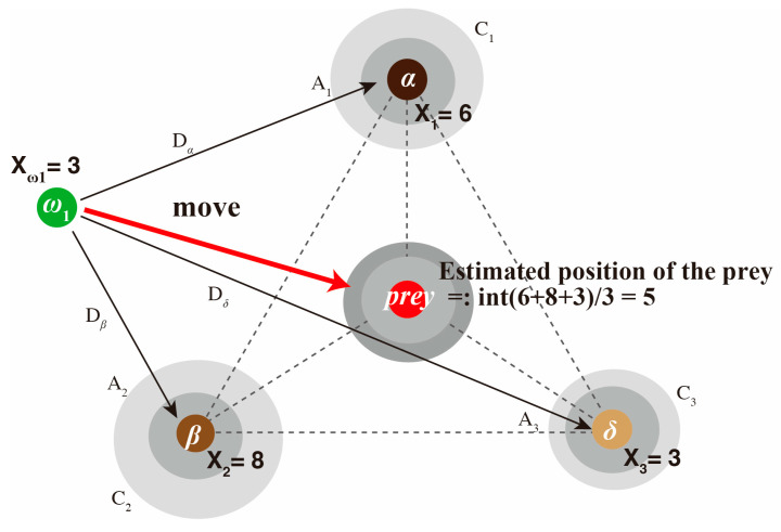
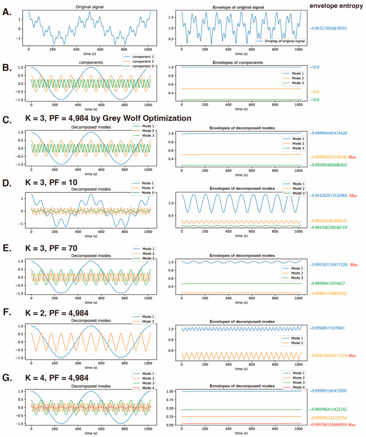
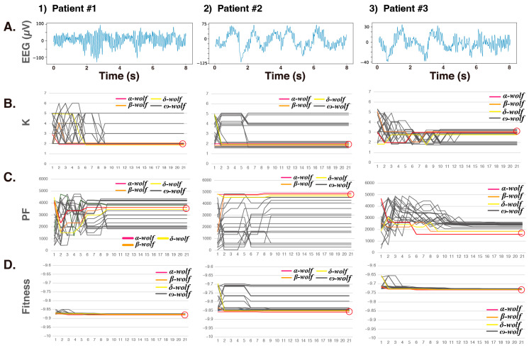
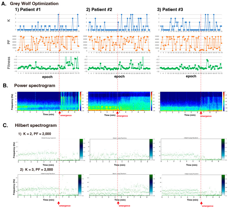
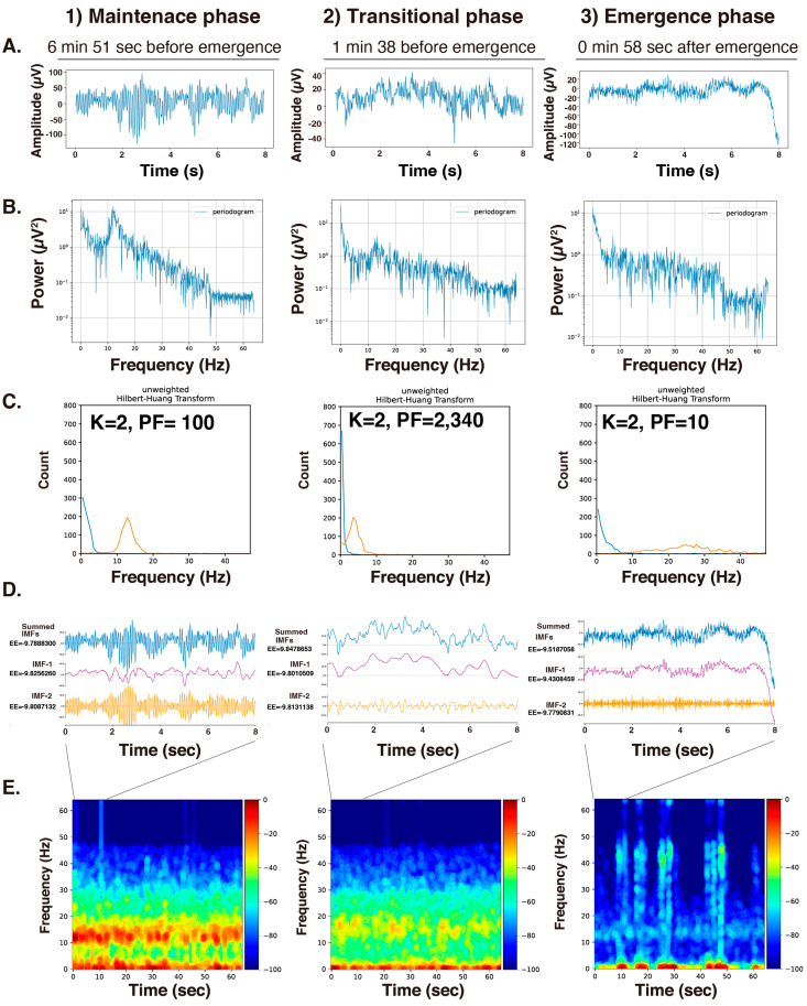
Frequency analysis via electroencephalography (EEG) during general anesthesia is used to develop techniques for measuring anesthesia depth. Variational mode decomposition (VMD) enables mathematical optimization methods to decompose EEG signals into natural number intrinsic mode functions with distinct narrow bands. However, the analysis requires the a priori determination of hyperparameters, including the decomposition number (K) and the penalty factor (PF). In the VMD analysis of EEGs derived from a noninterventional and noninvasive retrospective observational study, we adapted the grey wolf optimizer (GWO) to determine the K and PF hyperparameters of the VMD. As a metric for optimization, we calculated the envelope function of the IMF decomposed via the VMD method and used its envelope entropy as the fitness function. The K and PF values varied in each epoch, with one epoch being the analytical unit of EEG; however, the fitness values showed convergence at an early stage in the GWO algorithm. The K value was set to 2 to capture the α wave enhancement observed during the maintenance phase of general anesthesia in intrinsic mode function 2 (IMF-2). This study suggests that using the GWO to optimize VMD hyperparameters enables the construction of a robust analytical model for examining the EEG frequency characteristics involved in the effects of general anesthesia.

Keywords: Hilbert–Huang transform; electroencephalogram; general anesthesia; grey wolf optimizer; variational mode decomposition.


Conflict of interest statement

The authors declare no conflicts of interest.

Figures

Figure 1
Figure 1
Flowchart of grey wolf optimization (GWO) for variational mode decomposition (VMD).
Figure 2
Figure 2
Position updating algorithm for the wolves in the grey wolf optimizer. Three leader/subleader wolves, α, β, and δ, surround the prey, and the position of the prey is inferred from the positions of these three leader wolves, assuming that they surround it. Vectors A and C are coefficient vectors and are calculated for each coordinate. The other wolves in the pack, ω, locate the leader wolves’ positions, which are initially adjusted with a coefficient C and then gradually adjusted in each loop to better approximate the leader wolves’ positions. A random coefficient D is then applied to their distance, allowing them to approach the leader wolves within a range of −1 to +1. As a result, wolves that are closer to the prey than the leader wolves may replace them as the new leaders, enabling the pack to surround the prey more closely. The algorithm specifies three leader wolves and divides the obtained average position by the number of leaders. If the positions of α, β, and δ are 6, 8, and 3, respectively, then the position of the prey would be the midpoint, calculated as (6 + 8 + 3)/3 = 5.7, and the integer 5 becomes the updated position for the prey. The new position of the ω1 wolf is adjusted according to the leaders’ positions, taking into account its current position.
Figure 3
Figure 3
The relationship between the hyperparameters K and PF of VMD and the envelope entropy of each IMF. VMD is applied to a synthetic cosine wave composed of three known frequency components (2, 12, and 36 Hz). y=cos(2×2πt)+12cos(12×2πt)+14cos(36×2πt) (1024 data points). (A) The synthetic cosine wave, its envelope function, and the envelope entropy. (B) The three cosine waves, their envelope functions, and envelope entropies. (C) Decomposition of the synthetic cosine wave into three IMFs using the GWO algorithm with optimized values of K = 3 and PF = 4984, and calculation of each IMF’s envelope function and envelope entropy. (D) Application of VMD with K = 3 and PF = 10, and determination of IMFs, their envelope functions, and envelope entropies. (E) Application of VMD with K = 3 and PF = 70, and analysis of IMFs and their envelope functions, and envelope entropies. (F) Application of VMD with K = 2 and PF = 4984, and evaluation of the IMFs and their envelope functions, and envelope entropy. (G) Application of VMD with K = 4 and PF = 4984, and analysis of the IMFs and their envelope functions, and envelope entropies. K: decomposition number; PF: penalty factor; fitness: fitness value of VMD = the maximum envelope entropy of the IMF.
Figure 4
Figure 4
Optimization process for the hyperparameters K and PF of VMD using the GWO algorithm. Twenty wolves and 20 optimization loops, observed over one epoch (8 s, 1024 data points) of EEG during the maintenance phase of GA (obtained through continuous intravenous propofol administration) in three patients. This process involved monitoring the position of each wolf during two-dimensional wolf hunting. (A) EEG (8 s). (B) The optimization of K. (C) The optimization of PF. (D) The optimization of fitness. α wolf: red line; β wolf: orange line; δ wolf: yellow line; ω wolves: black lines. K: decomposition number; PF: penalty factor; fitness: fitness value of VMD = the maximum envelope entropy of the IMF.
Figure 5
Figure 5
Grey wolf optimization in the VMD of EEGs obtained from three patients of GA induced by continuous intravenous propofol. (A) The optimal solutions for K and PF were determined through fitness function optimization using the GWO algorithm. Twenty wolves and 20 optimization loops for all 73 epochs. Each epoch lasted for 8 s (128 Hz, 1024 data points) of the 10-minute period before and after awakening from GA. Additionally, K was set in the range of 2 to 6 (in increments of 1), and the PF values ranged from 10 to 5000 (in increments of 10). (B) The spectrogram for the 10-minute interval was obtained using the multitaper method and is displayed in Figure 4B. (C) Using VMD with K = 2 or 3 and PF = 2000, the signal was decomposed into IMFs and a Hilbert spectrogram was obtained. The gradient color bar shows the relative power value of the signal. K: decomposition number; PF: penalty factor; fitness: fitness value of VMD = the maximum envelope entropy of the IMF.
Figure 6
Figure 6
Analysis of 8 s EEG segments during the (1) maintenance, (2) transition, and (3) emergence phases of general anesthesia induced by propofol. (A) Original EEG (8 s, 128 Hz, 1024 data points). (B) The power spectrum obtained through Fourier analysis. (C) Hilbert spectrum. To optimize K and PF, GWO analysis was conducted with 20 wolves, 20 repetitions, and settings of K = 2 to 6 (in increments of 1) and PF = 10 to 5000 (in increments of 10). (D) Decomposed intrinsic mode functions (IMFs). (E) The power spectrogram for a 64 s period was determined using the multitaper method. The first 8 s were the subject of the VMD analysis. The gradient color bar shows the relative power value of the signal. K: decomposition number; PF: penalty factor; fitness: fitness value of VMD = the maximum envelope entropy of the IMF.

References

    1. Laferriere-Langlois P., Morisson L., Jeffries S., Duclos C., Espitalier F., Richebe P. Depth of anesthesia and nociception monitoring: Current state and vision for 2050. Anesth. Analg. 2024;138:295–307. doi: 10.1213/ANE.0000000000006860. - DOI - PubMed
    1. Roche D., Mahon P. Depth of anesthesia monitoring. Anesth. Clin. 2021;39:477–492. doi: 10.1016/j.anclin.2021.04.004. - DOI - PubMed
    1. Shander A., Lobel G.P., Mathews D.M. Brain monitoring and the depth of anesthesia: Another goldilocks dilemma. Anesth. Analg. 2018;126:705–709. doi: 10.1213/ANE.0000000000002383. - DOI - PubMed
    1. Chaddad A., Wu Y., Kateb R., Bourid A. Electroencephalography signal processing: A comprehensive review and analysis of methods and techniques. Sensors. 2023;23:6434. doi: 10.3390/s23146434. - DOI - PMC - PubMed
    1. Sharma R., Meena H.K. Emerging trends in EEG signal processing: A systematic review. SN Comput. Sci. 2024;5:415. doi: 10.1007/s42979-024-02773-w. - DOI

LinkOut - more resources