. 2024 Sep 13:15910199241282709.
doi: 10.1177/15910199241282709. Online ahead of print.

Perfecting the pour: A novel co-axial technique with sequential injections for optimising cement delivery during sacroplasty

Affiliations

Perfecting the pour: A novel co-axial technique with sequential injections for optimising cement delivery during sacroplasty

Mehran Nasralla et al. Interv Neuroradiol. .

Abstract

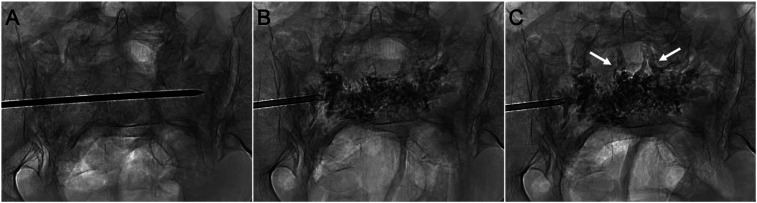
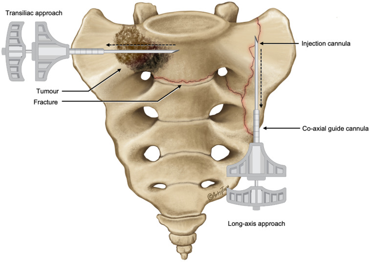
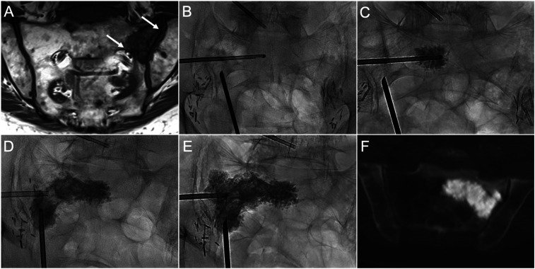
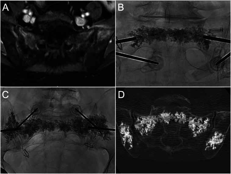
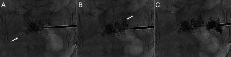
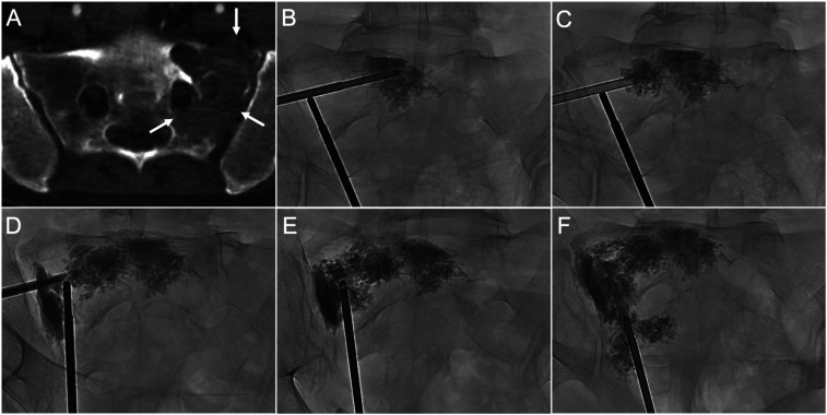
Background: Percutaneous sacroplasty is an effective treatment for painful sacral fractures and tumours, however there is no accepted optimal technique for performing this procedure. This study investigated a novel approach to sacroplasty combining co-axial sacral access, sequential cement injections and hypothermic cement manipulation to improve cement delivery.

Methods: This retrospective study analysed 11 patients who underwent co-axial sacroplasty between April 2023 and March 2024 for treatment of painful insufficiency fractures (n = 5) or malignant sacral tumours (n = 6). All cases were performed using biplane fluoroscopy with conebeam CT navigation for planning and monitoring percutaneous access. Procedural details, technical outcomes, and clinical outcomes including Numerical Rating Scale (NRS) pain and analgesic utilisation on a six-point scale were analysed pre-procedure and at follow-up.

Results: Technical success of was achieved in all cases using this technique. The mean injected cement volume was 20.5 ± 6.4 ml. Median pre-procedural NRS pain scores of 8 (IQR 7.25-8) significantly decreased to 0 (IQR, 0-0.25) at follow-up (p <.01). The median preprocedural analgesic utilisation score reduced from 3 (IQR, 2-3) to 0 (IQR, 0-2.5) at follow-up (p <.01). Cement leakage occurred during two cases without associated adverse clinical sequelae. There were no major adverse events.

Conclusion: Co-axial sequential injection sacroplasty is a safe and effective technique which allows facilitates controlled delivery of cement. Improved control of cement delivery, including around high-risk structures for cement leakage, offers a potential safety advantage over conventional sacroplasty techniques. Further research comparing technical and clinical outcomes to conventional techniques is warranted.

Keywords: Percutaneous sacroplasty; cement leakage; co-axial sacroplasty; sacral insufficiency fractures; sequential injection.


Figures

Figure 1.
Figure 1.
Epidural venous cement extravasation during conventional sacroplasty for bilateral sacral ala insufficiency fractures. (A) PA radiograph shows a transiliac approach at S1. The needle was inserted via the left sacral ala, traversing the sacral body to access the contralateral ala. (B) PA radiograph shows the cement distribution across the sacrum as the needle was withdrawn. (C) Epidural venous plexus extravasation (arrows) occurred following additional cement injections in the ipsilateral ala. This resulted from unintended cement propagation along the needle tract, rather than dispersing in the sacral ala around the needle tip.
Figure 2.
Figure 2.
Illustration of the positions of the outer co-axial guide and inner injection cannulas for transiliac and long-axis approaches. The outer guide cannula allows easy repositioning of the injection cannula across the target regions (dashed line arrows) to facilitate sequential cement injections while the guide cannula maintains sacral access between injections.
Figure 3.
Figure 3.
Co-axial sacroplasty of a left sacral ala metastasis. (A) Coronal T1 MRI image shows left ala metastatic lesion with erosions of the sacroiliac joint and S1 foraminal cortical margins (white arrows), which increase the risk of cement leakage. (B) PA fluoroscopic image demonstrates transiliac and long-axis guide cannula positioning. A side-opening injection cannula was inserted co-axially through the transiliac guide cannula across the lesion. (C) PA radiograph demonstrates the initial cement deposition. (D) Cement was injected through the long-axis guide cannula after initial sequential transiliac injections were completed. (E) Final distribution of cement after completion of long-axis and an additional transiliac injection in the ala superiorly. (F) Axial CT image shows contiguous cement distribution without evidence of foraminal or sacroiliac joint extravasation.
Figure 4.
Figure 4.
Co-axial sacroplasty and iliac cementoplasty for bilateral sacral and iliac insufficiency fractures, in a patient previously treated with pelvic radiotherapy for cervical cancer. (A) Axial T1FS with gadolinium demonstrates contrast enhancement in the sacral ala and iliac bones consistent with insufficiency fractures. (B) PA radiograph demonstrates cement distribution in the sacral body (Zone I) and periforaminal regions (Zone II) after sequential injections. (C) PA radiograph shows contiguous cement distribution from ala-to-ala at S1 following sequential cement injections, and bilateral iliac cementoplasties. (D) Axial CT image shows cement overall distribution and absence of cement leakage.
Figure 5.
Figure 5.
First application of the co-axial sequential injection technique involving a patient with insufficiency fractures involving the sacral body and right foramen. (A) PA radiograph after initial injection demonstrates cement in the sacral body and small presacral venous extravasation (arrow). (B) Subsequent injection around the foramen showed a small foraminal leakage (arrow). (C) Subsequent injection, following a period for cement curing, showed that the foraminal leak remained unchanged (arrow) without further propagation along the needle path. The foraminal leak was asymptomatic.
Figure 6.
Figure 6.
Application of side-opening injection cannulae in the treatment of complex sacral lesions. (A) Axial CT image demonstrates extensive osteolytic disruption of the left S1 foraminal, sacroiliac joint, and anterior sacral margins (arrows). (B–D) PA radiographs demonstrate sequential cement injections via the transiliac approach from the sacral body to the ipsilateral ala. (E–F) AP radiographs after long-axis sequential injections filled the remaining areas in the sacral ala to achieve a contiguous distribution. The side-opening was directed away from the foramen during cement injections. No foraminal, sacroiliac joint or extraosseous leakage was demonstrated.

References

    1. Lee J, Lee E, Lee JW, et al. Percutaneous sacroplasty: effectiveness and long-term outcome predictors. J Korean Neurosurg Soc 2020; 63: 747–756. - PMC - PubMed
    1. Kortman K, Ortiz O, Miller T, et al. Multicenter study to assess the efficacy and safety of sacroplasty in patients with osteoporotic sacral insufficiency fractures or pathologic sacral lesions. J NeuroIntervent Surg 2013; 5: 461–466. - PubMed
    1. M. Mok J, A. Strelzow J, L. Vallina V, et al. Anterior transpsoas approach for removal of cement leakage after sacroplasty. Spine Surg Relat Res 2022; 6: 563–568. - PMC - PubMed
    1. Tian Q-H, -Han K, Wang T, et al. Percutaneous sacroplasty with or without radiofrequency ablation for treatment of painful sacral metastases. American Journal of Neuroradiology 2022; 43: 1222–1227. - PMC - PubMed
    1. Frey ME, DePalma MJ, Cifu DX, et al. Percutaneous sacroplasty for osteoporotic sacral insufficiency fractures: a prospective, multicenter, observational pilot study. Spine J 2008; 8: 367–373. - PubMed

LinkOut - more resources