. 2025 Jan;297(1):105-119.
doi: 10.1111/jmi.13360. Epub 2024 Sep 14.

Setting up an institutional OMERO environment for bioimage data: Perspectives from both facility staff and users

Affiliations

Setting up an institutional OMERO environment for bioimage data: Perspectives from both facility staff and users

Anett Jannasch et al. J Microsc. 2025 Jan.

Abstract

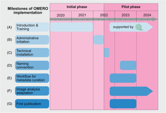
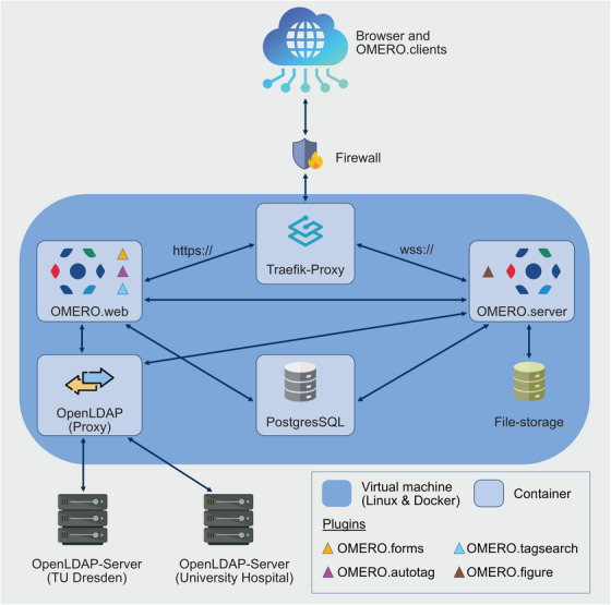
Modern bioimaging core facilities at research institutions are essential for managing and maintaining high-end instruments, providing training and support for researchers in experimental design, image acquisition and data analysis. An important task for these facilities is the professional management of complex multidimensional bioimaging data, which are often produced in large quantity and very different file formats. This article details the process that led to successfully implementing the OME Remote Objects system (OMERO) for bioimage-specific research data management (RDM) at the Core Facility Cellular Imaging (CFCI) at the Technische Universität Dresden (TU Dresden). Ensuring compliance with the FAIR (findable, accessible, interoperable, reusable) principles, we outline here the challenges that we faced in adapting data handling and storage to a new RDM system. These challenges included the introduction of a standardised group-specific naming convention, metadata curation with tagging and Key-Value pairs, and integration of existing image processing workflows. By sharing our experiences, this article aims to provide insights and recommendations for both individual researchers and educational institutions intending to implement OMERO as a management system for bioimaging data. We showcase how tailored decisions and structured approaches lead to successful outcomes in RDM practices. Lay description: Modern bioimaging facilities at research institutions are crucial for managing advanced equipment and supporting scientists in their research. These facilities help with designing experiments, capturing images, and analyzing data. One of their key tasks is organizing and managing large amounts of complex image data, which often comes in various file formats and are difficult to handle. This article explains how the Core Facility Cellular Imaging (CFCI) at Technische Universität Dresden successfully implemented a specialized system called OMERO. With this system it is possible to manage and organize bioimaging data sustainably in a way that they are findable, accessible, interoperable and reusable according the FAIR principles. We describe the practical implementation process on exemplary projects within scientific research and medical education. We discuss the challenges we faced, such as creating a standard way to name files, organizing important information about the images (known as metadata), and ensuring that existing image processing methods could work with the new system. By sharing our experience, we aim to offer practical advice and recommendations for other researchers and institutions interested in using OMERO for managing their bioimaging data. We highlight how careful planning and structured approaches can lead to successful data management practices, making it easier for researchers to store, access, and reuse their valuable data.

Keywords: FAIR principles; OMERO; bioimaging data; imaging facility; research data management (RDM).


Figures

FIGURE 1
FIGURE 1
Milestones of OMERO implementation. Illustration of the overall process of implementing OMERO at the CFCI during the initiation phase, with the focus on the administrative organisation and the pilot phase with the active usage of OMERO within different use cases. (A) Introduction and training covered two different periods: first, initial awareness and general education in the field of RDM and OMERO; and second, active practical training and learning after the installation of OMERO as actively supported by the I3D:bio team. (B) Six‐month phase of administrative processes. This was an important milestone as resource allocation enabled the start of the technical installation of the OMERO instance. (C) Completed over a period of 12 weeks challenges like accessibility of the OMERO instance within the medical domain and beyond as well as different user authentications were covered. Due to the group's lack of expertise in both RDM in general and OMERO specifically, multiple processes began in parallel. (D) Nine‐month development phase of a research group‐specific naming convention. This phase was necessary for closely linking the newly acquired skills to the in parallel‐developed workflows for metadata curation. (E) Focused period on understanding the tagging and developing of templates for Key–Value pair annotations. (F) Parallel integration of user‐specific data analysis processes from the beginning. This very time‐consuming process required specialised knowledge in image analysis and Python programming. This phase is still ongoing. (G) RDM of the data related to the PERIKLES project. Following a successful training and annotation phase, this project as part of the four pilot studies was the first one at the medical campus with figures created by OMERO.figure for a scientific publication that is now published.
FIGURE 2
FIGURE 2
Overview about the pilot projects. This figure was created in OMERO.figure as an example for FAIR‐bioimage publication. (A) Light microscopic image of a hematoxylin and eosin stained pericardial implant with granulocyte border area 1 week after subcutaneous implantation into a Sprague Dawley rat. (B) Lattice light‐sheet data used to study mitotic spindle scaling in the developing C. elegans embryo. The following components are labelled: cell membrane in magenta (PH‐domain labelled with mKate2), centrosomes in cyan (gamma‐tubulin labelled with GFP, arrows) and histones in magenta (histone H2B labelled with mCherry, arrow head). (C) Transmission electron microscopy image of a RPE‐1 cell in metaphase. Chromosomes can be seen in the centre of the cell as dark structures (arrows). One centriole pair is visible in the lower part of the cell (arrow head). (D) Light microscopic image of the parasite Loa loa in a blood smear stained with Giemsa staining. The characteristic structure microfilaria is indicated (arrow) and used within lectures and practical courses.
FIGURE 3
FIGURE 3
Technical OMERO setup. The Docker stack employed in this installation consisted of both, an OMERO.web (custom docker image) and an OMERO.server container (custom docker image), which are connected to a Traefik‐Proxy container (HTTP reverse proxy). Furthermore, the setup is constituted of containers for an OpenLDAP‐proxy (OpenLDAP custom image) and a PostgresSQL‐database. The OMERO.server container is a custom docker image, based on the image ‘openmicroscopy/omero‐server’ (hub.docker.com/omero‐server) and has been extended with the OMERO.figure plugin (github.com/omero‐figure). The OMERO.web container is also a custom docker image, based on ‘openmicroscopy/omero‐web‐standalone:5.22’ (hub.docker.com/omero‐web‐standalone), which has been extended with OMERO.forms (github.com/OMERO.forms), OMERO.autotag (github.com/omero‐autotag), and OMERO.tagsearch (https://github.com/German‐BioImaging/omero‐tagsearch). The user authentication is managed through the custom OPEN‐LDAP container. Since an LDAP integration was required for two domains, that is, for the Faculty of Medicine/University Hospital domain (MED) and the TU Dresden domain (ZIH), the OpenLDAP proxy was necessary to connect both domain trees.
FIGURE 4
FIGURE 4
Workflow for the ‘CountCellsOMERO’ macro. (A) Screenshot of the OMERO.web interface default viewer showing a representative image and the ROIs employed to select the area for image processing. For the displayed PERIKLES study, it was necessary to have an outline rectangle (dark blue) to ensure the mandatory size of the high‐power field (HPF; 1000 × 500 µm2 according to ISO 10993–6:2007 22 ). Here, one aspect of the project was to quantify cells within the fibrous capsule and implant matrix. Further segmentation as polygons (in green) within the rectangles were done to define both structures. (B) Screenshot of the Fiji dialog window shown at the beginning of the macro execution for parameter input. OMERO Login credentials, group and dataset ID needed to be selected. To choose polygons and not rectangles to be processed, we added the prefix term ‘batch_mask…’ and inserted this term in the dialog window. To process images of multiple datasets in a batch, we tagged all favored images with ‘to process’. (C) Screenshot of the OMERO.web interface showing the parameters and measurements from the macro saved as an attachment in OMERO. Parameters and measurements are displayed as a table (bottom left and right, respectively).

Similar articles

References

    1. Ferrando‐May, E. , Hartmann, H. , Reymann, J. , Ansari, N. , Utz, N. , Fried, H. U. , Kukat, C. , Peychl, J. , Liebig, C. , Terjung, S. , Laketa, V. , Sporbert, A. , Weidtkamp‐Peters, S. , Schauss, A. , Zuschratter, W. , & Avilov, S. (2016). Advanced light microscopy core facilities: Balancing service, science and career. Microscopy Research and Technique, 79, 463–479. 10.1002/jemt.22648 - DOI - PMC - PubMed
    1. Schmidt, C. , Boissonnet, T. , Dohle, J. , Bernhardt, K. , Ferrando‐May, E. , Wernet, T. , Nitschke, R. , Kunis, S. , & Weidtkamp‐Peters, S. (2024). A practical guide to bioimaging research data management in core facilities. Journal of Microscopy, 294, 350–371. 10.1111/jmi.13317 - DOI - PubMed
    1. Anderson, K. I. , Sanderson, J. , & Peychl, J. (2007). Design and function of a light‐microscopy facility. In Shorte S. L., & Frischknecht F. (Eds.), Imaging cellular and molecular biological functions (pp. 93–113). Springer Berlin Heidelberg. 10.1007/978-3-540-71331-9_4 - DOI
    1. Kemmer, I. , Keppler, A. , Serrano‐Solano, B. , Rybina, A. , Özdemir, B. , Bischof, J. , El Ghadraoui, A. , Eriksson, J. E. , & Mathur, A. (2023). Building a FAIR image data ecosystem for microscopy communities. Histochemistry and Cell Biology, 160, 199–209. 10.1007/s00418-023-02203-7 - DOI - PMC - PubMed
    1. Schmidt, C. , Hanne, J. , Moore, J. , Meesters, C. , Ferrando‐May, E. , & Weidtkamp‐Peters, S. (2022). Research data management for bioimaging: The 2021 NFDI4BIOIMAGE community survey. F1000Research, 11, 638. 10.12688/f1000research.121714.2 - DOI - PMC - PubMed

MeSH terms

LinkOut - more resources