. 2024 Oct;33(10):e5161.
doi: 10.1002/pro.5161.

Peptides inhibiting the assembly of monomeric human l-lactate dehydrogenase into catalytically active homotetramer decrease the synthesis of lactate in cultured cells

Affiliations

Peptides inhibiting the assembly of monomeric human l-lactate dehydrogenase into catalytically active homotetramer decrease the synthesis of lactate in cultured cells

Alessandra Stefan et al. Protein Sci. 2024 Oct.

Abstract

The energetic metabolism of cancer cells relies on a substantial commitment of pyruvate to the catalytic action of lactate-generating dehydrogenases. This coupling mainly depends on lactate dehydrogenase A (LDH-A), which is overexpressed in different types of cancers, and therefore represents an appealing therapeutic target. Taking into account that the activity of LDHs is exclusively exerted by their tetrameric forms, it was recently shown that peptides perturbing the monomers-to-tetramer assembly inhibit human LDH-A (hLDH-A). However, to identify these peptides, tetrameric hLDH-A was transiently exposed to strongly acidic conditions inducing its dissociation into monomers, which were tested as a target for peptides at low pH. Nevertheless, the availability of native monomeric hLDH-A would allow performing similar screenings under physiological conditions. Here we report on the unprecedented isolation of recombinant monomeric hLDH-A at neutral pH, and on its use to identify peptides inhibiting the assembly of the tetrameric enzyme. Remarkably, the GQNGISDL octapeptide, mimicking the 296-303 portion of hLDH-A C-terminal region, was observed to effectively inhibit the target enzyme. Moreover, by dissecting the action of this octapeptide, the cGQND cyclic tetrapeptide was found to act as the parental compound. Furthermore, we performed assays using MCF7 and BxPC3 cultured cells, exclusively expressing hLDH-A and hLDH-B, respectively. By means of these assays we detected a selective action of linear and cyclic GQND tetrapeptides, inhibiting lactate secretion in MCF7 cells only. Overall, our observations suggest that peptides mimicking the C-terminal region of hLDH-A effectively interfere with protein-protein interactions responsible for the assembly of the tetrameric enzyme.

Keywords: human lactate dehydrogenase A; inhibition of assembly; monomer; peptides; subunit–subunit interaction.


Conflict of interest statement

The authors declare no conflict of interest.

Figures
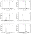
FIGURE 1
FIGURE 1
Isolation of monomeric and tetrameric human LDH‐A. Gel filtration of human LDH‐A previously subjected to affinity and hydrophobic interaction chromatography. The gel filtration chromatography was performed using: (a) 10 mM Tris–HCl; (b) 10 mM HEPES; (c) 10 mM Tris–HCl (black circles) or 10 mM Tris–HCl supplemented with 125 μM β‐NADH and 5 mM oxamate (white circles); (d) 10 mM HEPES, 150 mM NaCl; (e) 10 mM HEPES, 150 mM NaCl; (f) 10 mM Tris–HCl. A Superdex 200 column was used throughout, and all buffers were poised at pH 7.5. The white, cyan, red, magenta, blue, green, and black circles indicate the elution volume of ferritin, catalase, aldolase, albumin, ovalbumin, chymotrypsinogen, and RNase A, respectively. The absorption spectra of these molecular mass markers dissolved in Tris–HCl or HEPES buffer are reported in Figure S3.
FIGURE 2
FIGURE 2
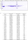
Analysis of monomeric human LDH‐A by SDS‐PAGE and mass spectrometry. (a) Electrophoretic analysis of fractions eluted from the Superdex 200 column equilibrated with 10 mM Tris–HCl (pH 7.5) and used to perform the last purification step of monomeric LDH‐A. M: Molecular mass markers (their M r is indicated in kDa at the left); I: Input; the fraction numbers are indicated on the top. (b) Identification by mass spectrometry of peptides obtained by in‐gel tryptic digestion of monomeric human LDH‐A.
FIGURE 3
FIGURE 3
Analysis of the quaternary and secondary structure of human LDH‐A. (a–c) Dynamic light scattering experiments performed with monomeric LDH‐A in: (a) 10 mM Tris–HCl, (b) 10 mM Tris–HCl supplemented with 125 μM β‐NADH, (c) 10 mM Tris–HCl containing 125 μM β‐NADH and 10 mM oxamate. All buffers were poised at pH 7.5. (d) Far‐UV CD spectra of tetrameric and monomeric LDH‐A (green and blue line, respectively), in PBS buffer.
FIGURE 4
FIGURE 4
Binding of β‐NADH to tetrameric and monomeric hLDH‐A. (a) Kinetics of the association of β‐NADH to tetrameric hLDH‐A as detected by surface plasmon resonance. Sensorgrams were observed loading increasing concentration of β‐NADH (0.49–125 μM) on a sensor chip modified with immobilized tetrameric human LDH‐A. (b) Determination of the K D of hLDH‐A for β‐NADH. The response units refer to the data shown in “a.” The continuous lines represent the best fit of a parametric rectangular hyperbola to the experimental observations. (c) Sensorgrams observed loading increasing concentration of β‐NADH (0.25–10 mM) on a sensor chip modified with immobilized monomeric human LDH‐A. (d) Linear dependence of the response units reported in “c” on the concentration of loaded β‐NADH. The continuous line represents the best fit of a linear equation to the experimental observations.
FIGURE 5
FIGURE 5
Kinetics of β‐NADH oxidation catalyzed by tetrameric or monomeric human LDH‐A. (a–c) Activity assays were performed using reaction mixtures containing 50 mM Tris–HCl (pH 7.5), 100 μM β‐NADH, and 500 μM pyruvate. (a) Kinetics of β‐NADH oxidation detected in the presence of 7.6 nM of tetrameric or monomeric LDH‐A (green and blue line, respectively). (b, c) Time‐course of β‐NADH oxidation catalyzed by 12.5, 25, 50, 100, or 200 pM tetrameric (b) or monomeric (c) human LDH‐A (magenta, cyan, red, blue, and green circles, respectively). The time‐course observed in the absence of enzyme is also reported (white circles). (d) Dependence on enzyme concentration of the rate constants determined for the reactions catalyzed by tetrameric (white circles) or monomeric LDH‐A (black circles) and reported in “b” and “c.” To obtain the k obs values a single‐exponential equation was fitted to the experimental observations.
FIGURE 6
FIGURE 6
Catalytic action of tetrameric and monomeric human LDH‐A. (a) Dependence of the initial velocity of β‐NADH oxidation observed as a function of pyruvate concentration, in the presence of 125 μM β‐NADH and 2.6 nM tetrameric (white circles) or 9.8 nM monomeric (black circles) human LDH‐A. The continuous lines represent the best fit of the Michaelis–Menten equation to the experimental observations. (b) Dependence of the initial velocity of β‐NADH oxidation observed as a function of β‐NADH concentration, in the presence of 500 μM pyruvate and 1.9 nM tetrameric (white circles) or 7.6 nM monomeric (black circles) human LDH‐A. The continuous lines represent the best fit of the Michaelis–Menten equation to the experimental observations. (c) Kinetic parameters determined by performing activity assays using monomeric or tetrameric LDH‐A, at the indicated final concentrations. Under conditions of variable pyruvate concentration, β‐NADH was always used at 125 μM. When reaction mixtures contained variable concentrations of the redox cofactor, pyruvate was invariably added at 500 μM. All the assays were performed at pH 7.5 (Tris‐BisTris, 10 mM each).
FIGURE 7
FIGURE 7
Design of the peptides to be tested against monomeric LDH‐A. (a) Details of the interactions between the N‐terminal region of one monomer with the residues 295–302 located at the C‐terminus of a second monomer, and (b) stick representation of the same region. Structural details of: (c) the G295QNG298 turn, and (d) the 13‐membered cyclopeptides c[GQN‐isoD] (TH2) and (e) c[GQN‐(R)‐isoD] (TH3), (f) the I299SDL302 turn, and the cyclopeptides (g) c[isoKSDL] (TH4) and (h) c[isoKSD‐(R)‐L] (TH5).
FIGURE 8
FIGURE 8
Inhibition exerted by linear and cyclic peptides on LDH‐A activity detected in vitro. Activity assays were performed with reaction mixtures containing 6.2 nM monomeric LDH‐A, 125 μM β‐NADH, and 500 μM pyruvate in Tris‐BisTris (10 mM each) buffer, pH 7.5 (white bar in “a”). The gray bars (in “a” and “d”) represent the activity observed in the presence of the same volume of DMSO (1%, v/v) carried to the assay mixtures by the peptides to be tested. (a–c) The effect, if any, of tetrameric peptides featuring the GQND primary structure (see also Table 1) on LDH‐A activity is shown. The inhibition exerted by the octameric peptide TH1 is also reported in “a.” The enzyme activity observed in the presence of linear or cyclic peptides is shown in “b” and “c”, respectively. (d) Extent of LDH‐A activity detected in the presence of linear or cyclic tetrapeptides featuring the KSDL primary structure. Error bars represent SD (n = 3). The experimental observations were compared by Student's t‐test. The ***, **, and * symbols denote p values lower than 0.001, 0.01, and 0.02, respectively. (e) Simulation of the association between the TH2 peptide and monomeric LDH‐A. Predicted interactions between the cyclopeptide c[GQN‐isoD] (TH2) and the N‐terminal sequence Y9NLLKEEQTPQ19, as simulated by molecular dynamics in a box of explicit TIP3P water molecules; gray and red dotted lines represent hydrogen bonds and salt bridges, respectively.
FIGURE 9
FIGURE 9
Effect of peptides on the secretion of lactate by human cell lines. (a) Relative levels of mRNA coding for LDH‐A or LDH‐B as detected by RT‐PCR in MCF7 cells. Only the mRNA coding for LDH‐B was detected in BxPC3 cells. (b) The amount of lactate secreted by MCF7 or BxPC3 cells in the absence (white bars) or in the presence (yellow, orange, and green bars) of different peptides is shown. Error bars represent SD (n = 3). The experimental observations were compared by two‐way ANOVA. The ** symbol denote a p value lower than 0.01.

Similar articles

References

    1. Arai K, Ishimitsu T, Fushinobu S, Uchikoba H, Matsuzawa H, Taguchi H. Active and inactive state structures of unliganded Lactobacillus casei allosteric l‐lactate dehydrogenase. Proteins. 2009;78:681–694. - PubMed
    1. Bernofsky C, Wanda SC. Formation of reduced nicotinamide adenine dinucleotide peroxide. J Biol Chem. 1982;257:6809–6817. - PubMed
    1. Bradford MM. A rapid and sensitive method for the quantitation of microgram quantities of protein utilizing the principle of protein‐dye binding. Anal Biochem. 1976;72:248–254. - PubMed
    1. Brooks GA. Lactate: glycolytic end product and oxidative substrate during sustained exercise in mammals: the lactate shuttle. In: Gilles R, editor. Circulation, respiration, and metabolism: current comparative approaches. Verlag, Berlin: Springer; 1985. p. 208–218.
    1. Chan WWC, Mosbach K. Effects of subunit interactions on the activity of lactate dehydrogenase studied in immobilized enzyme systems. Biochemistry. 1976;15:4215–4222. - PubMed

MeSH terms

LinkOut - more resources